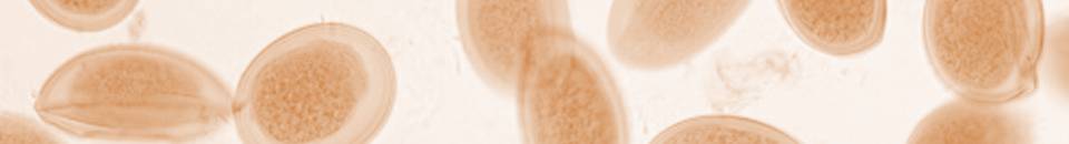
The diagnosis of intestinal helminthiasis in animals is most commonly performed indirectly by microscopy on faeces for the detection of the parasites’ eggs or cysts. This procedure involves suspending the sample in a special ‘Flotation Solution’ of an adjusted Specific Gravity (S.G. 1.20 – 1.27) which is higher than that of the eggs/cysts but lower than the other faecal debris which sink. This results in the eggs floating to the surface and which can then either be counted and/or identified by microscopy (x 100 magnification). The two types of method are described below:
Worm Egg Count Method
This method is useful for checking the worm burden of large, grazing animals (e.g. horses, cattle, sheep) which tend to have low worm burdens naturally. Animals shedding relatively large numbers of worm eggs in their faeces are at risk of loss of condition and illness and pose a threat to other animals grazing the same pasture. Worm egg counts (F.E.C.s) are also useful as part of a Faecal Egg Reduction Test to detect the efficacy of, or the resistance to, the anthelmintic in use. In general, F.E.C.s help vets and owners maintain their livestock in good health, provide financial benefits by avoiding unnecessary worming treatments and delay/avoiding anthelmintic resistance.
The McMaster Slide method of estimating the parasite load of an animal is made up of just four easy-to-understand and perform steps which can be viewed here
Performing the F.E.C. requires a Microscope, a McMaster Counting Slide, Flotation Solution andother small items ofequipment and consumables. However, especially for those who wish to start performing their own F.E.C.s and make it easier, we have bundled all these items together in discounted Bespoke DIY Kits, three of which include a microscope and one without.
Concentration/Flotation Method
This method is more appropriate for companion and small animals (e.g. dogs, cats) where the presence of any intestinal parasite is undesirable and so no egg count is necessary.
The basic principle uses a flotation solution of special Specific Gravity in which along with the faecal sample is placed in a special container with a coverslip placed on top touching the surface for 5 -10 minutes. After incubation the coverslip is removed vertically containing a drop of fluid underneath and placed on a microscope slide for examination. If any worm eggs/oocysts are present they will have floated to the surface and seen under the microscope
we would like to provide an update regarding ongoing market cost trends and their impact on our pricing.
Global market conditions have continued to drive sustained increases in direct costs, significantly affecting the entire supply chain. As a lot of the products supplied by Vetlab Supplies Ltd are polymer-based plastics derived from oil, fluctuations in oil prices and associated raw material costs have had a direct and considerable impact on production costs.
We have continued to make every effort to absorb these increases and minimise their effect on our customers, however, due to the ongoing nature of these issues, we have no option other than to implement an adjustment to our pricing & discounts on affected products from immediate effect.
Please note this will not apply to existing stock & will only be applied to new stock received at the increased price.
We would also like to emphasise that we will continue to closely monitor raw material and market cost developments. Should conditions improve, we will review our pricing accordingly and consider potential adjustments, including reductions where possible.