Ringworm is a dermophytic fungus that eats skin, hair, horn and claws in companion and commercial animals. Rapid veterinary diagnosis of dermophyte infection is the first step toward identifying, treating and eradicating fungal skin diseases.
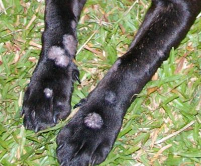
Dermatophytosis (ringworm) is distressing to pet owners. Not a worm at all, the classic bald patches and circles of reddened skin with flaking dandruff is caused by a fungus that feeds on the keratin protein of skin, hair and claws.
In cases of dermatophytosis, the usual suspects are the Microsporum fungi: M.canis, M.gypseum, M.persicolor, the Trichophytons: T.verrucosum, T.mentagrophytes and the Epidermophytons.
Ringworm infection is spread through skin flakes from infected animals and can be transmitted to humans. These flakes carry fungal spores called conidia. Conidia are resistant to antimicrobial treatments such as chloramphenicol and cycloheximide.
The size and shape of conidia enable the veterinary laboratory to identify the species of fungus. But first, vets need a fast and reliable diagnostic test to distinguish dermophytic fungal infection from other fungi and bacteria.
Mykodermoassay-Trio features three purpose-optimised fungal growth media
Mykodermoassay-Trio features three purpose-optimised fungal growth media on a single petri dish test-plate. Divided into three sectors, the time-saving diagnostic provides SAB/SDA, DTM and ESA media in separate agar-based fungal growth environments.
The Sabouraud (SAB/SDA) agar sector encourages fungal growth while subduing the growth of bacteria that might otherwise complicate interpretation. The gel’s optical clarity enhances visual inspection and macro-identification of fungal colonies.
The Dermatophyte Test Media (DTM) inhibits bacteria and non-dermophytes by the inclusion of chloramphenicol and cycloheximide. Phenol red pH indicator confirms any alkaline waste products characteristic of dermophytic fungi with an orange to red colour change.
Clarity enhances species identification
The Enhanced Sporulation Agar (ESA) sector, optimised for spore formation, includes the pH indicator bromophenol blue. A colour change from yellow to green-blue further supports the presence of dermophyte fungi. The gel’s clarity enhances species identification by veterinary microscopy.
Mykodermoassay-Trio provides for reliable interpretation in only 2 to 3 days. And performs equally well with samples of hair roots, dandruff, scabs and skin scrapings. Finally, a long fridge life of up to 24 months makes Mykodermoassay a convenient and cost-effective method for reassuring pet owners that their pet’s skin and fur will soon be off the menu for ringworm fungus.