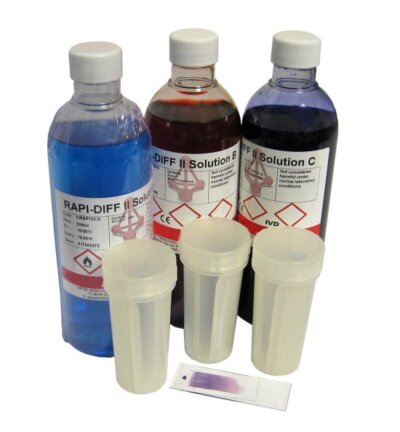
It’s an unfortunate fact that most things of interest in veterinary microscopy are all more or less colourless. Vetlab’s RAPI-DIFF II microscope slide staining kits help time-strapped veterinary laboratories make the invisible clearly visible.
Practically every living structure at the microscopic level exists in water and consists mostly of water. It’s so much a lack of colour that makes cells and cell structures difficult to see microscopically, it’s a lack of what veterinary microscopists call ‘contrast’.
Poor contrast leaves your subject no brighter or darker
Contrast is what makes an object of interest stand out from its background. In photos and TV images, poor contrast leaves your subject no brighter or darker than the less interesting stuff around, behind or even in front of it.
In a poor contrast picture, you don’t know where to look or even what you’re looking at. Poor contrast means disappointing holiday snaps, but could mean disaster when veterinary diagnosis depends on the micro-exam of a sample from your poorly pet.
Developing biological microscope stains – like dyes used in colour fabrics
Since the invention of microscopes in the 1590s, laboratories have worked to improve the contrast by developing biological microscope ‘stains’. Microscopic stains act just like the dyes used to colour fabrics.
Scientists found that some stains made particular microscopic structures stand out from the less interesting structures around them. Perhaps the two most interesting and important structures in veterinary microscopy are:
- The Cytoplasm. This is all the stuff that’s inside the cell membrane. Staining the cytoplasm makes the cells stand out and easier to see. It’s also easier to determine the shape of cells which helps diagnose some parasitic infections
- The Nucleus. This is the cell’s central control unit that contains the DNA. Many cells, especially infection-fighting white blood cells, have particular shaped nuclei. Identifying white cell types can help vets diagnose some diseases.
In 1891 the Russian physician Dmitri Leonidovich Romanowsky invented a balance of microscope stains for cell cytoplasm and nuclei that not only made cells easier to find and examine, but actually helped identify different kinds of cell.
Romanowsky’s proven staining principle
Vetlab’s RAPI-DIFF II kit incorporates Romanowsky’s proven staining principle into a complete, convenient and simple to use microscope slide staining kit.
Pre-prepared solutions create a simple three–dip process that fixes the sample to the microscope slide, stains cell cytoplasm with a Thiazine-Eosin Y, and contrasts clearly with the Methylene Blue stained nuclei. Selectively rinsing with pH buffered solution or further dipping gives veterinary labs a practical tool for recognising and differentiating a range of cell types and diagnosing abnormalities.