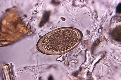
McMaster Slide Test: Starting Out in DIY Animal Parasite Load Testing

McMaster Slide Test: Starting Out in DIY Animal Parasite Load Testing

The restrictions imposed during the worst of the Covid-19 pandemic forced keepers of pet, domestic and rescued animals to carry out their own parasite load monitoring using the McMaster Slide Faecal Egg Count (FEC) test.

With veterinary practices and diagnostics mostly back to normal, many keepers continue to easily and cost effectively monitor their own animals. Here’s how to get started with only the most essential of veterinary laboratory equipment and an easy to follow McMaster Slide Counting Method.

Get Started In DIY Faecal Egg Monitoring

The McMaster Slide method of estimating the parasite load of an animal is made up of just four easy to understand and perform steps.

First, separate the worm eggs from a sample of the animal’s faeces. Second, find the eggs (oocytes) using a microscope and, third, count them. Finally, knowing both the weight of faeces sampled and the number of worm eggs counted, calculate the animal’s likely level of worm infection.

Egg Counting: What You Need – Flotation Solutions

Not as obvious as the microscope and McMaster counting slide, the Flotation Solutions are the key to the success of egg counting and identification. Flotation solutions are made to a standard or customer specified, specific gravity (SG) more usually known as density.

The fluid density is chosen such that faecal matter and other debris is more dense than the solution and so sinks. But the parasite eggs are less dense, and so float to the top making finding, counting and identifying them straightforward with the right equipment.

Egg Counting: What You Need – The Microscope

Essential to starting out on faecal egg monitoring is a microscope capable of ranging from 40-times (x40) to 100-times (x100) magnification. The Vetlab Premiere Range of microscopes are economical, easy to use and popular with diagnostic and teaching laboratories.

The microscope will be used to find and count the number of parasite eggs present in a small sample of animal faeces. With a bit of practice it’ll become straightforward not only to estimate the number of eggs, but even identify the likely species of worm causing infection.

Egg Counting: What You Need – The McMaster Counting Slide

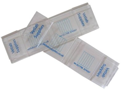
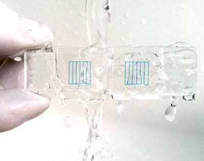
The McMaster Counting Slide is the simple yet brilliantly adapted microscope slide used and relied upon by animal health practitioners since 1939. Made in glass or tough acrylic plastic, the slide is basically the carrier for a square cavity of known volume – usually 0.15ml.

After a sample of faeces, treated with a specific ‘flotation solution’ is placed in the McMaster Slide cavity and covered with a second, thinner slide. Parallel lines etched into this ‘cover slide’ create five equal divisions. Viewed with the microscope, the floating parasite eggs – just under the cover slip – can be counted and identified within each division.

Egg Counting: What You Need – The Simple Final Calculation

With a little practice, counting the number of parasite eggs seen within the grid lines of the McMaster Slide becomes a quick and easy routine.
As long as the Vetlab McMaster Slide Counting Method has been followed precisely, all that remains is to multiply the counted number of parasite eggs by 25 to get the number of eggs per gram (e.p.g.) of animal faeces.
For more information on the Vetlab McMaster Slide Count Method, veterinary laboratory equipment, ready-made or customised flotation solutions, search or click Vetlab F.E.C  Kits & Equipment


For further information visit our website www.vetlabsupplies.co.uk or call 01798 874567

Hedgehogs to Llamas: The McMaster DIY Faecal Egg Count

Hedgehogs to Llamas: The McMaster DIY Faecal Egg Count

Simple, reliable and cost effective, the McMaster slide Faecal Egg Count (FEC) test equips you to monitor the parasitic load and intestinal health of your pet, commercial and rescue animals.

Left untreated, parasitic intestinal worms in animals from hedgehogs to llamas and alpacas can cause serious illness and even death to infected animals.

A wide variety of parasitic worms can reproduce inside loved and valuable animals. Infection spreads through millions of microscopic eggs shed into the faeces of even one host animal to contaminate shared bedding, stabling, pasture or foodstuff.

Impact Of Covid 19 On Routine Faecal Parasite Egg Monitoring

For pet, commercial and rescue animal keepers, restricted access to routine veterinary monitoring was one of many distressing side effects of the Covid 19 pandemic.
Managing the risks to animal health, even by such readily treatable conditions as parasitic intestinal worms, became much more critical in the absence of regular and reliable testing.

Do It Yourself Diagnostic Faecal Egg Counting (FEC) Techniques

Conveniently, for pet, commercial and rescue animal keepers, the veterinary standard diagnostic for intestinal worms can now be simply, reliably, and economically carried out as a DIY in-house test.
Although microscopic parasitic intestinal worms can be difficult to observe and identify, their eggs – and their potential to cause significant further infection – are larger and more easily observed and identified.

Identifying Intestinal Worms And Estimating Parasitic Load

Identifying and estimating an animal’s ‘parasitic load’ uses pre-prepared solutions and basic laboratory equipment, readily available from Vetlab Supplies. Vetlab’s easy-to-follow laboratory method guides you through the taking of samples, preparing the test, examination under the microscope and identifying the type and likely numbers of parasitic intestinal worms.
The straightforward method is known to veterinary surgeons as the McMaster Slide FEC (Faecal Egg Count) Test. A laboratory standard since 1939, The McMaster Slide is basically a small glass cavity of known volume with a calibrated transparent cover that can be viewed under a microscope.

The McMaster Slide Standard Laboratory Faecal Egg Count (FEC)

The McMaster Slide Test relies on the simple fact that, in specified flotation solutions, parasite worm eggs float, while most of the other solid matter in animal faeces tends to sink. So if the number of parasite eggs in a measured weight of faeces can be counted, then the parasitic load in the animal’s digestive system can be easily calculated.
And, because the eggs of different parasites have different characteristics, so the careful use of a veterinary microscope and a guided choice of specific flotation solutions, the type and species of intestinal worm can be identified for relevant treatment and control.

Identify Parasites With Off The Shelf Or Custom Made Flotation Solutions

Vetlab Supplies offers a range of off the shelf and customised faecal flotation solutions ideal for general and species specific egg count testing. Matching the density (specific gravity) of the flotation solution to the density of the target worm species eggs allows a qualitative as well as quantitative estimate of an animal’s parasitic load.

For McMaster Slides manufactured in glass and robust acrylic, together with all necessary flotation solutions, as well as economical easy to set up and use microscopes, just click or search Vetlab Supplies F.E.C Kits & Equipment


For further information visit our website www.vetlabsupplies.co.uk or call 01798 874567

UriStain Urine Sediment Staining

UriStain Urine Sediment Staining

Aids Diagnosis of Kidney Disease

Sediment in centrifuged animal urine samples generally indicates some sort of renal malfunction. UriStain™ urine sediment staining and microscopic examination provide a convenient method for identifying cellular and non-cellular sedimentary components and contributing to a timely, accurate diagnosis.

Clouded Urine Is Obvious, But The Cause Might Be More Obscure

Healthy kidneys generally produce clear, cloudless urine with little or no insoluble matter. Urine with a murky appearance, caused by suspended solid material, could indicate kidney malfunction potentially leading to complete renal failure.

Kidney failure may already be suspected based on other observed symptoms or reports of incidents such as injury or poisoning. Abnormally opaque or coloured urine samples can provide veterinary laboratories with a diagnostic opportunity without the need for invasive tissue sampling biopsy.

Centrifugation, Staining And Microscopic Examination Of Urine Sediment

Centrifugation of a urine sample in a veterinary centrifuge will cause any solid matter to concentrate at the bottom of the sample tube. Microscopic examination of the re-suspended precipitate may assist in identifying the type and source of any solid material.

Unambiguous visualisation and differentiation of the sedimentary components are essential to the rapid and accurate diagnosis of the cause of any renal problem. Hardy Diagnostics UriStain from Vetlab Supplies is formulated to enable veterinary laboratories to categorise the most likely solid materials responsible for clouding in animal urine samples.

Clear Differential Staining Of Cellular And Non-cellular Components

Colour responsive UriStain™ assists the veterinary microscopist in distinguishing between living cellular material, and non-viable cells, cellular fragments and other non-soluble material. Parallel examination of an unstained microscopic preparation helps identify sedimentary components based on their unstained as well as on their stained morphology.

Red blood cells, in suspensions of urinary sediment, will stain faintly pink, while the nuclei of white cells and other epithelial cells appear a deep purple colour. Yeast cells, recognisable by their morphology, also appear purple as do dead bacteria. Fungal mycelia and spores show a lighter purple. The parasitic protozoan, Trichomonas, stains light blue but might also appear colourless.

Fat droplets, with their characteristic ‘honeycomb’ appearance, remain unstained. Together with their morphology, hyaline casts – a mucoprotein potentially indicative of glomerulonephritis, and other granular material, can also be characterised by their response to UriStain visualisation.

Ready To Use And No Filtration Required

Composed of certified source dyes, the UriStain balance of Ammonium Oxalate, Safranin and Crystal Violet provides veterinary laboratories with a stable, convenient and practical staining protocol based on a reformulation of the Sternheimer-Malbin urine sediment procedure.

Ready to use, the UriStain™ reagent is supplied in convenient 15ml dropper bottles and requires no filtration prior to adding to the centrifuged urinary sediment. Veterinary microscopes equipped with low power magnification (100X) and high power magnifications (400X) enable the stained components to be precisely identified and quickly cell-counted if required. This product is directly comparable to Sedi-Stain, if not better!

For further information visit our website or Tel: 01798 874567 and we will be delighted to help.


Brucellosis Vaccine

Brucellosis Vaccine

Initiative Calls For New Collaborative Approach

The Global Alliance for Livestock Veterinary Medicines (GALVMed) is calling for veterinary laboratory, academic and industrial collaboration toward ‘milestone two’ in developing and manufacturing an effective vaccine against the livestock disease, brucellosis.

Brucellosis causes abortions, infertility, lower milk production

Caused by infection with the bacterium Brucella melitensis. Brucellosis causes abortions, infertility, lower milk production and weight loss among cows, sheep, goats and buffalo herds. Brucellosis is ‘zoonotic’, meaning that it can, under some circumstances, cross the species boundary and infect humans. The annual £359m economic burden of brucellosis falls most heavily on small scale and subsistence livestock keepers in the developing countries of South Asia and Sub-Saharan Africa.

Symptoms of brucellosis include swollen udders, swollen testicles in males, nervousness, fever and, most characteristically premature abortion. At present, there is no effective vaccine against B. melitensis, a situation that the Ag Med managed ‘Brucellosis Challenge’ means to correct.

Combating brucella infection

The difficulty in combating Brucella infection rests on its ability to escape the attention of the host animal’s immune system. Even though it doesn’t form resistant spores, the aerobic, gram-negative coccobacilli can survive and multiply within the very cells designed to destroy them. Safe within these ineffective killer-cells, the host’s circulatory system carries the invader into the central nervous, genital-urinary, pulmonary and musculoskeletal system.

The ‘Brucellosis Challenge’ is a funded by AgResults – on behalf of the Australian, Canadian, UK and the US governments, and the Bill and Melinda Gates Foundation. The 10 year project incentivises participating veterinary laboratories, academic institutions and industrial manufacturers with ‘milestone’ funding prizes each worth up to £70,000.

Three ‘milestones’ (develop an idea, prove the concept, register the product) mark the progress of the challenge toward its goal. With ‘milestone two’ in sight, the aim is to produce a brucellosis vaccine that is: safe for pregnant animals, effective in more than 80% of vaccinated animals, deliverable in a single, annual vaccination all at an affordable cost and with a long shelf life.

Testing for Brucella canis

Long shelf life and affordability are key features of the FASTest Brucella Canis test for the causative organism of brucellosis in dogs. B. Canis causes abortion typically after 45-55 days although, because dead foetuses might be reabsorbed, the observed symptom might be a failure to conceive. In males, the main symptom may be testicular degeneration and semen with no sperm, reduced sperm or large numbers of abnormal sperm resulting in infertility. Non-specific signs in both dogs and bitches include lethargy and apparent premature ageing.

Requiring no refrigerated storage and delivering a clear-cut colour-change result in 20 minutes, the cost-effective diagnostic veterinary test is a true field test for use on site at kennels and canine breeders.

To find out more about our large range of veterinary diagnostic test kits visit our website: www.vetlabsupplies.co.uk or Telephone: 01798 874567

Lower Lambing Season Losses

Lower Lambing Season Losses

Fast Field Test for E.coli- K99 Pathogen

The first lambs of the lambing season are here and full of the joys of the spring yet to arrive. Delivering as early as the second week in January, the most productive ewes will give birth to twins or even triplets, with quadruplets not uncommon and the occasional delivery of quintuplets.

Large numbers of new-borns fail to thrive

Sadly, the arrival of so many newborn lambs over a relatively short time span can result in a large number of new-borns failing to thrive early, sicken or die. In well managed flocks, mortality among newborn lambs is reported to be as low as 5%, but loss rates can be as high as 20% in extreme circumstances. Every lamb lost is damaging to the economic survival of Britain’s highly pressured sheep farms.

High mortality among newborn commercial animals

One all too common cause of high mortality among newborn commercial animals is pathogenic E.coli infection. A particularly pathogenic strain of the common gut bacteria, Escherichia coli, is known in veterinary laboratories as E.coli-K99. The K99 tag refers to a molecular feature on the surface of the E.coli bacteria that differentiates it from other, less harmful strains. The feature effectively disguises the invader from the host animal’s protective system of search-and-destroy ‘phagocyte’ cells that normally seek out and engulf unwelcome bacteria rendering them harmless. Unrecognised by infection-fighting phagocytes, the multiplying bacteria infiltrate the host animal’s digestive system causing severe tissue damage and resulting in dangerous fluid loss through uncontrollable diarrhoea.

Identifying pathogenic E.coli infection

Veterinary laboratories have exploited the K99 marker as a means of identifying pathogenic E.coli infection, allowing treatment with antibiotics that can also identify, target and destroy bacteria with the K99 antigen.

The ability to quickly and confidently identify K99 E.coli is vital because severe diarrhoea and dehydration is deadly not only in newborn lambs, but also in calves and piglets. Diarrhoea in newborn animals can have multiple causes, including infection with enterotoxic Rotavirus, Coronavirus, Cryptosporidium, Salmonella and Giardia, so it can be difficult to make a quick diagnosis based on symptoms alone.

Rapid and reliable – all-in-one veterinary field test

Vetlab Supplies FASTest E.coli-K99 Strip is the genuine go-anywhere veterinary field-test for E.coli-K99 in lamb, calf and piglet faeces. Rapid and reliable, the all-in-one kit requires no refrigerated storage or laboratory equipment to produce a quick, clear-cut, colour change diagnosis of K99 infection.

FASTest E.coli-K99 Strip, and other Vetlab Supplies veterinary diagnostic kits provide timely and reliable support to the veterinary examination of animals suffering pathogenic digestive tract infections.

To find out more about our large range of veterinary diagnostic test kits visit our website: www.vetlabsupplies.co.uk or Telephone: 01798 874567

Survey of Horses and Owners Shows Importance of FEC Monitoring

Survey of Horses and Owners Shows Importance of FEC Monitoring

A survey of horse owners at the 2017 Royal Welsh Show found that better awareness of routine faecal egg counting (FEC) in horses could reduce the need for veterinary treatment.

The UK’s county-based agricultural shows are an ideal opportunity for producers, consumers and their suppliers to gather and exchange information toward the improvement animal husbandry, health and welfare.

Unique opportunity to gather information

The Royal Welsh Show, held each July at the Llanelwedd showground in Builth Wells, Powys is among the largest of the county agricultural shows both in the UK and in Europe. The gathering of so many from so far afield provides veterinary laboratory researchers with a unique opportunity to gather real-world information from a wide range of animals and their owners.

At the 2017 Royal Welsh Show, researchers from the Institute of Biological, Environmental and Rural Sciences at Aberystwyth collected faecal samples from 60 horses taking part in the four-day event. Using a commercial faecal egg flotation system they recorded the number of nematode eggs per gram (epg) of faecal matter, and related their findings to monitoring and treatment details supplied by horse owners.

Faecal egg counting in horses at the show revealed detectable nematode infection in 30 (50%) of tests. Almost a third (27%) were scored above the 200 epg baseline generally considered as requiring anti-parasite treatment. Questioning the owners and keepers revealed that, on average, horses received 3 anthelmintic treatments per year, though some received none, and others as many as 6.

18% of owners confessed to irregular FEC testing

Asked about the frequency of faecal egg count analysis, almost a fifth (18%) of owners confessed to irregular and infrequent testing, while only two of the sixty reported a routine approach with repeat testing every eight weeks. The value of FEC monitoring was reflected in the fact that only two of the regularly tested horses returned egg counts above the 200 epg threshold.

Overall, the average egg count of untested horses was more than four times that of animals whose owners committed to routine faecal egg monitoring. The benefit of regular FEC testing was further proven by the researchers’ observation that horses not subject to routine FEC analysis were more than 150 per cent more likely to require veterinary treatment.

While the survey strongly suggests that owners are not over-medicating their horses, it also indicates a low level of FEC monitoring in the wider horse-owning community. Researchers from the Institute of Biological, Environmental and Rural Sciences hope that the Royal Welsh Show findings will highlight the crucial role of routine faecal egg counting in horses in equine health and welfare management.

To find out more about our large range of veterinary products visit our website: www.vetlabsupplies.co.uk or Telephone: 01798 874567

Paramphistomiasis In The UK – Accurate Egg Counting Is No Fluke

Paramphistomiasis In The UK – Accurate Egg Counting Is No Fluke

Paramphistomiasis, or rumen fluke, is of increasing interest as a parasitic disease of livestock producing a range of non-specific symptoms including diarrhoea, weight loss and general weakening.

Paramphistomes are two-host trematode parasites that spread their lifecycle between mammals and molluscs. Grazing on infected snail material introduces parasite cysts into a ruminant’s digestive system where hatched-out flukes feed before heading up into the rumen. Here they attach to the rumen wall and feed on its contents like a mass of pink maggots. Resistant eggs (oocytes) pass through the gut and out with the host’s faeces. Taken into roving snails, the oocysts hatch and reproduce to complete the life cycle.

Observed in British livestock as far back as the 1950s

Rumen thrives in the warm moist tropical climate with little significant impact on livestock economics. More recently, the organism has become increasingly common in temperate climes infected an estimated 20% to 30% of European livestock. Recent studies report the parasite as present in up to 77% of sheep in Ireland with prevalence across the UK varying between 29% and 52%.
Observed in British livestock as far back as the 1950s, the prominent paramphistome species identified in the UK is Calicophoron daubneyi. Since the late 2000s, the trematode’s eggs have started appearing in routine Veterinary Investigation Diagnosis Analysis (VIDA) examinations.

Detecting the heavier eggs

Rumen fluke eggs are relatively heavy, compared to the oocysts of most intestinal parasites. Consequently, centrifugation using flotation solutions and convenient recovery systems such as Ovatube are not efficient in the detection of rumen fluke oocysts in animal faeces. Veterinary laboratories generally employ ‘sedimentation’ rather than ‘flotation’ techniques to detect the heavier eggs of trematode flukes.

In sedimentation, a small sample of faeces is thoroughly suspended in water and the bulk of solid material removed using a coarse metal or cloth strainer. Left to settle, the sediment from the filtrate is then re-suspended in clean water. Adding a drop of methylene blue or malachite green to the recovered solid material from this suspension will clearly stain remaining faecal material blue or green, leaving parasite eggs unstained.

Diagnosis and control of this new invader

A veterinary microscope equipped with 10x eyepieces and a 10x objective allows identification of Paramphistomum and other relatively heavy parasite oocysts including Fasciola hepatica (liver fluke). Using a McMaster Worm Egg Counting Slide, to relate the number of eggs found to the number of faeces sampled, allows a quantitative assessment of the level of parasite infection.

The likely economic impact of the spread of rumen fluke is as yet uncertain. Fortunately, because its eggs can be detected during examinations for liver fluke in faecal samples, and its similar life cycle, veterinary laboratories are forewarned and forearmed in the diagnosis and control of this new invader.

To find out more about our large range of veterinary products visit our website: www.vetlabsupplies.co.uk or Telephone: 01798 874567

Egg Counts and Coccidians – Controlling Coccidiosis in Cattle and Sheep

Egg Counts and Coccidians – Controlling Coccidiosis in Cattle and Sheep

Coccidiosis is an intestinal disease caused by single-celled coccidian parasites. Though there are several species of coccidian protozoa, all must get inside the cells lining the intestines of their host to reproduce.

Coccidians spread when their eggs (oocysts) shed with their host’s faeces contaminating the food of other potential hosts. Mainly associated with poultry, infected birds suffer enteritis with blood stained diarrhoea, becoming lethargic, anaemic and showing a generally degraded condition.

Distinguishing between coccidiosis and similar symptoms

Poultry coccidians don’t infect farm mammals, nor do the cow and sheep equivalents infect chickens and waterfowl. In birds, veterinary microscopy of tissue from the characteristically swollen intestines of confirms coccidian infection. In sheep and cows, distinguishing between coccidiosis and similar symptoms of colibacilliosis, cryptospiridiosis, coronovirus, rotavirus and bovine viral diarrhoea (BVD) can require further veterinary laboratory investigation.

Veterinary diagnostic kits equip busy vets with a fast and reliable answer to the question of which parasite is responsible for the observed symptoms. Vetlab’s FASTest kits provide accurate, early diagnosis even in the field allowing treatment and preventative measures to begin immediately.

Using histological staining techniques

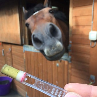
Where coccidiosis is indicated, faecal oocyst counts can give an estimate of the level of infection. Faecal egg count flotation solutions, Ovatube detection kits and smooth, quiet veterinary centrifuges make oocyst recovery quick and clean. Veterinary microscopes and histological staining techniques support the quantitation and identification of particular coccidian species.

In recent years, histological and egg count surveys have estimated the coccidian infection rate of cattle to be about 20%. However, not all species of coccidians cause disease, and a heavy oocyte burden doesn’t always indicate a clinically significant infection.

The risk is especially high where many animals are confined

Sever coccidiosis in calves and lambs can result in life-threatening dehydration. Infection usually follows from ingestion of oocyte-infected faeces or contaminated food. The risk is especially high where many animals are confined in faeces-soiled enclosures or where young animals are grazed on land contaminated by material from hosting adults.

The exception to coccidian species specificity is Toxoplasma gondii. T.gondii reaches maturity and reproduces only in cats, where it causes more serious symptoms. Toxoplasmosis only rarely causes illness in humans and generally only in immunologically weakened dogs.

T.gondii oocysts in faeces from roaming cats can initiate abortion or foetal reabsorption in sheep. The veterinary response to this commercial risk includes vaccination, husbandry and animal-health expertise with quick detection and diagnosis with the FASTest Toxoplasmosis g diagnostic kit.

To find out more about our large range of veterinary diagnostic test kits visit our website: www.vetlabsupplies.co.uk or Telephone: 01798 874567

Resistance Is Useless: Antimicrobials– Awareness, Behaviour and Diagnostics

Resistance Is Useless: Antimicrobials– Awareness, Behaviour and Diagnostics

Super-bug resistance to antimicrobial treatment is a serious problem for pig, poultry and cattle farmers in the UK and worldwide. Many antibiotics ‘of last resort’ are longer effective against infections caused by bacteria such as Klebsiella pneumoniae and Escherichia coli.

Antibiotic resistance is set to cost the world’s commercial farmers an estimated 10 million animal deaths by 2050. So said the UK’s Chief Veterinary Officer reporting to The London Vet Show last November. That compares to a current figure of about 700,000. Yet the potential global cost could be tackled at only a fraction of the cost in money and suffering.

Exposure to ineffective levels of antimicrobials

Inappropriate and incomplete courses of antimicrobials in the past is blamed for a large part of the problems vets face today. Exposure to ineffective levels of antimicrobials kills only the most vulnerable pathogens leaving resistant bugs to survive and pass their protection to their offspring.

Over-use of antimicrobials has the same effect. Because microbes have a short generation time, there’s a high chance of a random resistant mutation occurring in an environment already dosed-up with antimicrobials. Again, all subsequent generations will have in-built resistance.

Improved public awareness and legislation

Defeating AMR involves improved public awareness and legislation to change treatment behaviour as well as research into new drugs. Proposed actions include 10-year targets to reduce unnecessary usage together with restrictions on the most crucial antimicrobials. Improved transparency allows the public to support antimicrobial-free food production practices through their shopping choices.

Veterinary laboratories can reduce the need for antimicrobial treatments with quicker and more specific veterinary diagnostics. Many bacterial infections cause a similar range of symptoms in infected animals. Diagnostics that can differentiate between causative organisms help to identify the most appropriate treatments and dosage, reducing the opportunity for new resistance to arise.

Laboratory scientists found that even resistant superbugs have an Achilles heel

Faced with using just the current arsenal of antimicrobials, researchers at the Ross University School of Veterinary Medicine on the island of St Kitt’s, report all is not lost. Laboratory scientists found that even resistant superbugs have an Achilles heel. They found that both Klebsiella pneumoniae and Escherichia coli each had a gene in their DNA that could be turned off. Once these genes were deactivated, the bugs lost their resistant super-powers and were once again vulnerable to the drugs previously used to fight against them.

As the scientific battlefront against AMR pushes forward, evidence suggests that the fight to change behaviour is at least holding its ground. Results from the European Surveillance of Veterinary Antimicrobial Consumption show a decline in antimicrobial usage of 2.4% 2011-2014. This downward trend supported by improving veterinary diagnostics means that for super-bugs, resistance may yet be useless.

Rinderpest Eradication – A Veterinary Success Story

Rinderpest Eradication – A Veterinary Success Story

On 8 May 1980, the World Health Organisation declared the devastating human disease, smallpox, officially eradicated in every country of the world. Only one other disease has been similarly eradicated at the global level: the 2000-year-old disease of wild and domesticated cattle, Rinderpest.

Similar to the measles and canine distemper virus, rinderpest is a single-stranded RNA paramyxovirus invading via the respiratory tract through contact with infected body fluids or contaminated drinking water.

The first outbreaks were recorded in Asia as far back as the 4th century

Rinderpest symptoms include fever, mouth ulcers, diarrhoea and mucosal congestion followed by a general weakening and death within 8 to 12 days. Also called steppe-morain and cattle-plague, the disease is almost 100% lethal in domestic herds of cattle, buffalo, yak and wild cloven-hoofed animals such as wildebeest and giraffe.

The first outbreaks were recorded in Asia as far back as the 4th century and first properly investigated in the 17th. The earliest experiments in immunisation began in the 18th century in response to economic collapse and starvation following the devastating waves of cattle plague that swept relentlessly through Asia, Africa and Europe.

The virulent nature of the disease is exemplified by the severe epidemic that took hold in Europe following the transit of Indian cattle through Antwerp en route for Brazil in 1920. The outbreak led directly to the formation of the Office International des Epizooites (OIE) now known as the World Organisation for Animal Health.

The OIE began a programme of global rinderpest eradication

The OIE began a programme of global rinderpest eradication beginning first with improved animal hygiene, husbandry and disease containment until the first production in 1957 of Walter Plowright’s effective vaccine.

In 1999, the OIE predicted the global eradication of rinderpest by 2011. Despite occasional outbreaks in the decade that followed, in 2011, the rinderpest paramyxovirus was declared as only the second disease, after human smallpox, to be officially eradicated world-wide.

The OIE maintains a post-eradication role of monitoring and control

Although no longer present in live animals, virus stocks are still held in about 20 laboratories around the world. Though intended for vaccine production, the risk of an accident or an act of bio-terrorism remains; and the OIE maintains a post-eradication role of monitoring and control of virus storage facilities.

Eradication programmes for other animal diseases are likely to follow. As with rinderpest, the strategy is likely to involve not just the search for a vaccine, but also the rigorous application of high standards in hygiene, husbandry and containment, together with advances in veterinary laboratory practice, sample management, examination techniques and diagnostic testing.