Spring In The Beehive – Time For A Bee Health Check?

Spring In The Beehive – Time For A Bee Health Check?

Spring In The Beehive – Time For A Bee Health Check?

Spring weather boosts honeybee activity and with it the risk of disease and colony collapse. Here’s a fast test to help hobbyist and commercial beekeepers keep a check on colony health.

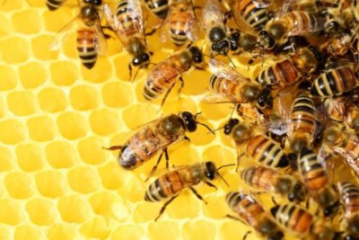
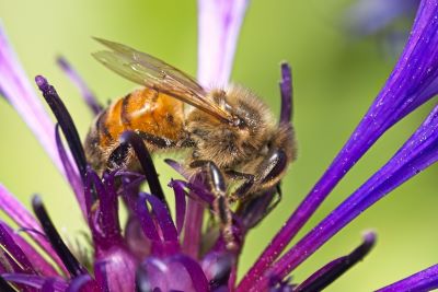
It’s spring! And whether they know it or not, garden fruit growers and farmers will owe almost all of their future harvests to the growing number of enthusiast and commercial honeybee keepers.

Spring Is Here And The Bees Are Getting Busy

These early months of warmer and dryer weather, together with the increasing day length, give honeybees the boost they need to get out there and get busy.

Just as spring is time for the honeybees themselves to get active, March, April and May are months for the enthusiast, hobbyist and commercial beekeeper to really get involved in the seasonal monitoring of hive and colony health.

Know The Risks To Your Bees In Springtime

Vital as these fascinating and seemingly aerodynamically impossible little flyers are to human food security, they’re really only looking to their own growth and survival through the winter to come.

Knowing the risks ahead, and getting prepared for potentially devastating problems, is the best approach to maintaining the health of adult bees and their broods through spring and summer in preparation for autumn and winter.

Spring In the Beehive Month By Month

As March progresses, your honeybee colony will be growing quickly and might need a sugary food boost. If a check while cleaning the floor tray shows low in-hive reserves, a natural bee pollen paste might fill the energy gap and support the health of the emerging colony.

By April, the occasional drone (with its larger head and much bigger eyes) might appear as broods increase and colonies grow quickly. Now is the time to identify the queen and maybe block the queen from laying eggs in the honey ‘supers’ to ensure a future harvest of clean honey. The potential for swarming could also become a risk in the stronger hives.

Through May, the queen is laying across most of the brood box as nectar and pollen come in thick and fast. This is the time for some honey harvesting especially near oil seed rape fields, and for adding more ‘supers’. Weekly inspections will help you be ready to capture any swarming bees into another hive.

Increasing Warmth Increases The Risk Of Disease

Increasing hive activity brings increased risks to honeybee health as rising temperatures encourage predators and disease carrying parasites to become more active and more numerous.

This is why the spring months maybe the best time for enthusiast and commercial honey producers to get ahead of the growing risk by testing and responding quickly to results that indicate a present or potential health risk.

Mite Spotting: The All Important First Check

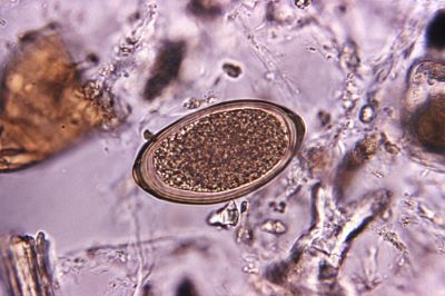
Knowing how to search out the 1.1mm to 1.7mm shell-shaped mite makes them easy to spot and count. Without the distinct body ‘segments’ typical of ants and spiders, the tell-tale shiny red-brown pinhead bodies and cluster of tiny legs help characterise Varroa destructor.

Methods for finding and counting the mites in a busy, active colony – including Natural Varroa Drop and Brood Uncapping – were tried and tested on the Advanced Beekeeping Course run by the National Diploma of Beekeeping (NDB) at Pershore College in Worcestershire.

One of their exercises was to compare different methods of monitoring the number of Varroa mites in a colony. Best-selling beekeeping magazine and international award winner, BeeCraft were invited to observe the process and its results. You can read about the Varroa project methods and results on the BeeCraft Varroa Webpage here.

Just What Does The Varroa Mite Do?

The UK Government Animal and Plant Health Agency (APHA) National Bee Unit summarises Varroa destructor as a parasite that feeds on the growing honeybee larva’s equivalent of the human liver. In their active months, adult worker bees can interact with neighbouring colonies spreading mites and worsening infestations.

The parasite drastically reduces brood resistance to a range of viruses including Deformed Wing Virus (DWV). The DWV virus either kills pupating larvae outright or, if they do emerge as adult bees, they show deformed wings and die within two to three days.

Varroa mites multiply on worker and drone larvae inside their brood cells and escape on the adult’s body as it leaves the chamber. High levels of spring and summer mite infestation can increase the risk of winter colony collapse where the majority of worker bees disappear leaving a hive that cannot sustain its queen and brood cells.

Risks of Over Treating Hives for Varroa

The National Bee Unit Best Practice Guidelines describe two types mite killing varroacides which, if used appropriately, can be effective at reducing Varroa mite infestation.

Single-action synthetic pesticides have a single, specific mode of action. Specificity brings with it the high risk of resistance leading to the reduced effectiveness appearing in the UK and Europe.

Non-synthetic pesticides have multiple actions which reduce the risk of resistance. While the NBU website recommends these ‘natural pesticides’ over synthetics, it also instructs that all applied varroacidal treatments should be recorded.

The National Bee Unit recommends treating for Varroa in all colonies of an apiary at the same time in order to reduce the risk of resistance and colony reinfection to and from nearby hives.

Monitoring And Recording Varroa Infestation

The Animal and Plant Health Agency (APHA) National Bee Unit specialises in supporting hobbyist and commercial beekeepers. The NBU online publication Managing Varroa names the spring and early summer months as key to monitoring Varroa mite infestation.

Monitoring Varroa mite infestation, and effective testing for the potentially devastating diseases they carry, can save yours and other keepers’ colonies from expensive and increasingly ineffective antiviral treatments.

Testing Your Hives Reduces Overtreatment and Resistance

Spring and early summer testing for the paralysing ABPV (Acute Bee Paralysis Virus) and the malformed-wing and body-weakening DWV (Deformed Wing Virus) before any treatment. This approach will help reduce virus resistance and might save your entire colony when infection strikes.

For more detailed information on the symptoms, effects and treatment of these viruses simply click this link to our ‘Health Check Your Honey Bees’ page.

Simple, Fast Effective: The 3-in-1 DIY Bee Health Check

Professional veterinary laboratories use a DNA test known as the polymerase chain reaction (PCR), to confirm varroa-borne infections. This is not a practical option for hobbyist, small scale and most other bee keeping enthusiasts. Fortunately, a reliable and easy to use diagnostic test kit is available ‘off the shelf’ which can give early warning of mite borne CBPV and DWV risks to hive health.

The Vetlab Supplies FASTest BEE 3T is the quick and easy diagnostic check for Deformed Wing Virus (DWV) and Acute Bee Paralysis Virus (ABPV) and includes an additional, third test, for the colony destroying Sac Brood Virus (SBV). SBV reduces larvae from healthy white to sickly yellow, then to a shrivelled dark brown or black ‘Gondola’ or ‘Chinese Slipper’ curved shape.

The Changing, Challenging World For Honeybees

In recent years, global warming has led to increased average and maximum temperatures in the UK and Ireland. Increasing hot-weather highs threaten instant, though short lived, crises for honeybee keepers due to the effects of heat and drought on bees, hives and food sources.

Rising average temperatures pose a more long-lasting danger as plant food sources fail to adapt rapidly enough to a hotter more arid environment. Worse still, predators and parasites better survive milder winters and become more active earlier in the new season.

Fighting these environmental ills may be beyond the means of beekeepers. But with an understanding of the challenge, a range of treatments which can prove effective and the pre-emptive advantage offered by modern diagnostic tests, the battle is anything but lost.


For further information on FASTestBee 3T and on the full range of animal health diagnostic kits please visit  www.vetlabsupplies.co.uk or telephone 01798 874657

Count Your Faecal Parasite Worm Eggs Before They Hatch

Count Your Faecal Parasite Worm Eggs Before They Hatch

Counting parasite worm eggs in the faeces from your animals is easy and economical with Vetlab Bespoke FEC starter kits, that include flotation solutions, veterinary quality microscopes and everything you need to carry out Faecal Egg Counts.

Internal parasites of grazing animals spread from one animal to another by infective eggs shed with the faeces of an infected animal. Parasite eggs hatch into the grazing pasture where the active worms spread out and are eaten by grazing animals repeating the cycle of infection and increasing the ‘worm burden’ of all exposed animals.

Worm eggs, passed out in the faeces of infected animals, can lie dormant through the winter to reinfect grazing animals when they return to pasture in the spring. Heavy worm burdens reduce growth rate, cause a loss of weight, body condition and increase vulnerability to infection resulting in emaciation and even death.

What Is A Faecal Egg Count?

A faecal egg count or FEC is a measure of the number of parasite worm eggs in an animal’s faeces (droppings). Results, presented as the number of worm eggs per gram (epg) of faeces, gives an indication of the level of infection or ‘worm burden’ in an individual animal or for the entire herd.

The Faecal Egg Count is a useful, non-invasive method for estimating the individual worm burden of grazing animals including horses, cattle, donkeys, sheep, goats, llamas and alpacas. Owners of a small number of animals may be able to check every individual animal regularly, while larger herds or commercial farms may rely on representative sampling rather than each individual animal.

Why Are Regular Faecal Egg Counts Necessary?

Zero or medically insignificant Faecal Egg Counts will reassure keepers as to the internal health of their animals and to the hygiene and cleanliness of their pasture and stables. Confirming that no medical intervention is necessary will save on costs, and ensure that any veterinary treatment required is targeted where it will bring most benefit.

Medicating animals only when necessary prevents the overuse of drug treatments. In recent years, the ready availability of anti-worm medicines has led to a ‘treat first, test… maybe’ approach by some keepers. Overexposure to unnecessary medication has led to increasing drug resistance in parasitic worms and the growing risk of all available treatments becoming ineffective.

How Often Should An FEC Be Carried Out?

In the UK, Vettimes reports that FEC testing should be performed in all horses at 8-12 week intervals throughout the grazing period (spring to early autumn). Testing should also be carried out at the end of winter if horses have been grazed outdoors frequently or for longer periods.

Alpaca and llama specialists, The British Alpaca Society recommends advice from your veterinarian as permissible levels of FEC may depend on the age/condition of the animal and the type of worm. As a general rule, says BAS-UK, it is common to treat far lower egg counts in alpacas than might be considered in other animals.

For Sheep, the industry led Sustainable Control of Parasites in Sheep (SCOPS) recommends regular FEC testing on a 2 to 4 weekly cycle throughout the grazing season. SCOPS recognises that collecting faecal samples from every animal in a commercial flock isn’t practical!

This SCOPS link tells you how best to get a reliable overview of the parasite burden in a large flock. Collecting fresh droppings from a representative number of animals for professional laboratory or DIY testing is suggested as a testing strategy.

Can I Carry Out Faecal Egg Counts On My Own Animals?

Collecting fresh faecal samples from your horses, cattle, donkeys, sheep, goats, llamas and alpacas is the first step in reliable DIY faecal egg counting. SCOPS recommends wearing gloves to collect faeces less than 1 hour old and washing hands thoroughly afterwards. For large flocks, sampling 10% of the animals is advised.

Samples can be sent to commercial veterinary laboratories for testing but, with a little practice and some basic laboratory equipment, owners and keepers can carry out their own DIY in-house FEC testing to support their vet in deciding if any further treatment is necessary.

What Laboratory Equipment And Supplies Do I Need for FEC Testing?

How much DIY faecal worm egg counting you choose to do yourself depends only on your level of self confidence, readiness to gain experience and access to the appropriate and readily available laboratory equipment and consumables.

The FEC process divides readily into four stages: recovery of appropriate faecal samples, parasite egg recovery, egg counting, and interpretation of results. Depending on those results, a fifth stage might involve any appropriate veterinary treatment and any necessary changes to pasturing, stabling and hygiene practice.

How Can I Start DIY Animal Faeces Sampling?

SCOPS recommends collecting a representative number of fresh (warm to the touch) faecal samples from your animals. Disposable gloves and hand cleanser are also recommended as the minimum level of protective hygiene. Everything you need to carry out your own hygienic sampling can be found on the Vetlab Supplies Laboratory Consumables pages

What Do I Need To Separate Parasite Eggs from Faeces?

Parasite eggs are less dense than the faecal material that contains them. Left for long enough, a dispersion of faeces in water would allow the eggs to float free of the faecal material. However, this process is too slow to be practical, so gravity needs a little help to speed things along.

Mixing the faeces with a solution that encourages the eggs to float and the faecal matter to sink is made easy with Vetlab Supplies Ready-Made and Bespoke Floatation Solutions.

How Do I Count The Number Of Worm Eggs In A Faeces Sample?

Counting worm eggs sounds like a difficult task. But with a few pieces of the right kit, and a little practice, keepers can become proficient at estimating the worm burden of their animals. This count will equip keepers with sufficient information to approach their vet for advice on the appropriate anti-worm treatment.

Parasite egg counting requires one of the easy to use and economically available microscopes as recommended by Vetlab Supplies. The Vetlab helpline can guide you through the process of selecting and obtaining a microscope appropriate to your need.

Counting parasite eggs is made easy by the use of the tried and tested McMaster Worm Egg Counting Slide. The McMaster Slide holds a fixed volume of the liquid from the faeces sample. The number of worm eggs in this volume of sample can be counted by viewing the slide under the microscope.

All of the equipment required is available to purchase individually or as part of a complete bespoke kit.

How Do I Know If My Animals Need Veterinary Treatment?

A simple maths formula, calculated from how much of the faecal sample was tested, how much flotation solution was added, and how many eggs were counted on the McMaster slide gives you and your vet a measure of the worm burden for the animal tested.

With this FEC knowledge, you will be able to discuss with your vet the best course of any required treatment and helping prevent the emergence of multi-resistant worm infections in your animals.

To find out more about our range of Bespoke Parasitology Starter kits and the Vetlab McMaster Worm Egg Counting Method visit our website: www.vetlabsupplies.co.uk or call Tel: 01798 874567

Simple Equine Tapeworm Testing: Straight From The Horses Mouth

Simple Equine Tapeworm Testing: Straight From The Horses Mouth

Regular testing for equine tapeworm in horses and donkeys with the Equisal Saliva Test and Bespoke FEC Kits can increase the effectiveness of anthelmintic treatments, reduce the risk of parasite resistance and promote the long-term health of your horses and donkeys.

Overtreatment Increases Parasite Resistance

Treatment with equine de-worming medicines, known as anthelmintics (AH), effectively controls the vast majority of intestinal parasitic worms (helminths). However, the ready availability of these same medicines has lead to their overuse as a quick and easy substitute for veterinary testing and livery stable hygiene.

Tolerance to AH drug treatments in parasitic worm populations ranges from ‘highly susceptible’ to ‘highly resistant’. Overexposure to AH treatment might kill the vast majority of parasites, but the more resistant worms – though initially fewer in number – may still survive and multiply.

Without competition from the more susceptible worms, the fewer highly resistant survivors thrive, breed and pass on their drug-resistance to the next generation. The outcome is that overmedication reduces the effectiveness of each AH treatment until no useful treatment option remains.

Stronger and Stronger Doses Are Not The Answer

Increasing anthelmintic resistance (AHR) means that even stronger doses of AH medicines are needed next treatment. Continued over-treatment with the same AH medicine again and again only strengthens the resistance of the surviving tapeworms.

Eventually, the treatment becomes ineffective or, worse still, only effective in a dose so strong as to risk a dangerously adverse reaction from the horse itself. Changing the drug of choice may only begin a further round of acquired resistance ending in yet another ineffective treatment.

Strategies To Avoid Acquired Drug Resistance

Constantly changing the AH regime means that the few survivors of one treatment are less likely to survive a second, different, treatment before further resistance develops. With twice-yearly laboratory testing and good stable hygiene, unnecessary and ineffective treatment can be avoided.

With no new anthelmintic treatment on the horizon, the future relies on changes in the behaviour of owners and keepers. New strategies to counter increasing drug resistance in digestive tract parasites include regular monitoring and treating medically only when necessary.

Testing before treating not only helps prevent the development of drug-resistant tapeworms, it reduces the cost of wasteful over-treatment. The British Horse Society recommends a planned timetable of spring and autumn testing with medication, only when justified by the test results.

Good Hygiene Is Key to Equine Health

The need to resort to new treatment strategies is, perhaps, an indication that previous parasite reduction regimes have not been effectively applied. A report by UK-VET EQUINE (2009), circulated by The Horse Trust identifies the key risk factors in the spread of AHR, including:

– Poor manure collection and cleaning regimes.
– Poor pasture and paddock management.
– Frequently changing and high density horse populations.
– Grazing of younger, more vulnerable horses with older animals.
– Lack of effective quarantine measures.

UK-VET EQUINE recommended a range of effective strategies to reduce these risk factors:

– Collection of faecal material at least twice weekly.
– Rest and rotation of pasture – especially on stud farms.
– An awareness that parasites can overwinter on pasture.
– An awareness that worm larvae can migrate into pasture from contaminated field edges.

Types of Equine Intestinal Worm Infections

Horses and donkeys are at risk of infection and reinfection from a range of intestinal parasitic worms collectively known as Helminths. These worms fall broadly into three groups classified by veterinary laboratories as flukes (trematodes), tapeworms (cestodes) and roundworms (nematodes).

Animal charity Blue Cross warns that animals kept in poor, overstocked or unhygienic conditions are vulnerable to serious illness caused by five subtypes of intestinal parasitic worm:

Large Redworms (strongyles). Living in the horse’s intestine, they cause a swollen abdomen, internal bleeding, colic, weight loss and diarrhoea.

Small Redworms (cyathostomins). Feed on the intestinal tissue, these worms burrow into the gut wall to lie dormant through winter months emerging in early spring to cause weight loss, loss of condition, distended abdomen, colic and diarrhoea.

Roundworms (ascarids). Growing up to 40cm long, roundworms exploit the immature immune systems of foals under four years to cause inflammation and obstruction resulting in poor growth, lethargy, coughing, weight loss, loss of condition, distension and colic.

Tapeworms (anoplocephela). Tapeworms attach to the gut wall at the junction of the small intestine and large intestine causing impaction, colic, weight loss and even physical damage.
Tapeworm infestation is a particular problem for horse and donkey keepers because tapeworm eggs are too small for detection by routine centrifugation and McMaster slide FEC methodology.

Assessing The Worm Burden: Faecal Egg Counting

Equipped with a veterinary microscope, flotation solutions and a McMaster counting slide , the tell-tale eggs of most intestinal worms can be separated from horse faecal material. Faecal egg counts can accurately estimate the degree of any worm burden.

The tale-tale eggs of equine tapeworm are too small as to be accurately recovered and counted using the tried and tested methods of centrifugation and McMaster faecal egg counting. Detection of equine tapeworm infestation requires a new and innovative veterinary test.

Simple Saliva-Swab Test for Tapeworm in Horses
EquiSal Tapeworm Saliva Testing Kit for Horses is an Enzyme Linked Immunosorbant Assay test known as ELISA. The Equisal ELISA test searches out any antibodies produced by a horse’s immune system in response to tapeworm infection.

The EquiSal Tapeworm test works like an antibody blood test but uses saliva which horse
owners can collect themselves using a specially designed mouth swab as shown in this video . The swab is then simply packed in the preservative provided in the kit and posted to the laboratory in the freepost bag provided.

How Good Is The Equisal Saliva Tapeworm Test?

In a 2016 large-scale stables trial of Equisal, only 22% of the 749 horse tested were found to require veterinary treatment. This means that 78% – 583 horses – might have been unnecessarily and expensively treated if Equisal testing had not been carried out.

The economics of Equisal testing speak for themselves. Targetted treatment is not only economical, it also reduces the frightening prospect of runaway drug resistance resulting in debilitating and potentially lethal equine tapeworm infections.

To Treat or Not to Treat – That is The Question

The only 100% accurate way to measure a horse’s tapeworm burden would be count the number of worms within the horse’s intestines. In validation studies, the EquiSal Tapeworm test only misreported horses as tapeworm free when fewer than 20 tapeworms were present.

Parasitologists consider horses and donkeys showing fewer than 20 tapeworms to have a non-pathogenic medical condition. This means that these tests confirm the EquiSal Tapeworm Test as a success in identifying all pathologically significant tapeworm burdens.

Vets and stable owner can find all their technical questions about Equisal Testing answered in detail here, on Vetlab’s EquiSal Tapeworm FAQs page.

Check Your Rescue Dogs for Canine Brucellosis

Check Your Rescue Dogs for Canine Brucellosis

Testing re-homed and rescue dogs for Canine Brucellosis is simple with this in house testing kit.

Canine Brucellosis is on the rise for kennel keepers, puppy breeders and pet owners. Here’s why the simple and effective FASTest Brucella Canis test is now the vital off-the-shelf resource for dog rescuers, breeders and re-homers.

Rising Brucellosis Risk From Imported Rescue Dogs

The recent confirmed case of dog-to-human Brucella infection matches the steep rise in imports of rescue dogs from brucellosis endemic countries. Veterinary professionals, dog breeders and animal charities are voicing their concerns for the health of untested UK dogs and their human carers.

Human Brucellosis Risk From Untested Dogs

Before the Covid lockdown of 2020, there were just 3 UK cases of canine Brucella infection. This shot up with 97 more cases in just 6 months mostly traceable to imports or contacts with dogs from Eastern Europe. Human Animal Infections and Risk Surveillance (HAIRS) assesses the risk to human health from Brucella Canis as generally very low. But the risk may be higher for breeders of imported dogs, vets spaying and neutering infected dogs and anyone with a weak or suppressed immune system.

Symptoms and Treatment for Canine Brucellosis

Infected dogs may show tiredness and swollen lymph glands as well as stiffness and lameness. Brucella Canis infected dogs often suffer infertility and miscarriages. People exposed to Brucella Canis can suffer fever, headaches, tiredness and joint pain together with loss of appetite and weight. Most recover with antibiotics, but infection can lead to endocarditis or even meningitis if left untreated.

Confirmed Rescue-Dog to Human Brucella Infection

Imports of dogs from Belarus, Poland, Romania and Ukraine were temporarily halted after a confirmed dog-to-human Brucella Canis infection in August 2022. The BBC reported that Mrs Hayes of Stoke-on-Trent was hospitalised with constant shivering, severe headaches and severe back pain. Diagnostic tests showed that Mrs Hayes had contracted canine brucellosis. Moosha, Mrs Hayes’ recently re-homed Belarus rescue dog and most likely source of infection, also tested positive for Brucella Canis. Mrs Hayes recovered after antibiotic treatment, though her five dogs had to be destroyed to halt the threat of infection spreading more widely.

Link Between Brucellosis And Dog Imports

Most of the 11 to 12 million dogs in the UK live in domestic households with 31% housing more than one or more dog. 2023 DEFRA data shows a big increase in commercially imported dogs, including rescue dogs, rising from 41,313 in 2018 to 78,299 in 2021. Vet Times estimated that imported dogs make up 8% of the UK canine population. Of the veterinary professionals surveyed, Vet Times reported that 61% had seen increases in exotic diseases such as Brucella Canis in just two years. In April 2021, positive tests for Brucella Canis in dogs became legally reportable to the Animal and Plant Health Agency (APHA) brucella group, and to the DAERA Direct Regional Office in Northern Ireland.

Vets and Pet Charities Call for Testing

Brucella Canis is a ‘zoonotic’ infection, meaning that under some conditions it can pass from one species of animal to another. People with weakened immune systems, a prior illness or close and sustained contact with infected dogs are at greatest risk. Animal charities such as the PDSA are advising that dogs travelling from abroad should be tested for brucellosis before entering the UK. The British Veterinary Association (BVA), urges a proactive approach to reduce the Brucella Canis risk before, rather than after, dogs arrive in the UK. The Royal Veterinary College recommends testing for all imported dogs, dogs with a travel record to Brucella Canis endemic countries, and for any dog that has clinical signs that might indicate Brucella infection.

Safety For Rescue And Re-homed Dogs

UK Government advises that rescued and re-homed animals, and breeding stock not previously spayed or castrated, represent the highest risk of infection. Rescuers and prospective owners are urged to test for Brucella Canis a month before importation and to have the animals neutered where possible. Ensuring dogs are Brucella free prior to arrival protects UK dogs and people as well as avoiding potential veterinary costs. Any dog previously in contact with a dog from a Brucella Canis endemic country should also be tested. Dogs suspected of infection should be kept away from other dogs and people until testing is complete. Suspected infections should be made known to a veterinary surgeon, who can contact the Animal and Plant Health Agency (APHA) for further information and advice.

Keep Your Pets and Yourself Biosecure

The risk to human health from B.Canis is considered low. However, minimal contact with reproductive or birthing products, blood, and urine is advised. Washing hands thoroughly for a minimum of 20 seconds with soap and hot water, or the use alcohol gel sanitiser after handling such materials, is also strongly recommended. If B.canis symptoms are suspected, you should contact your GP or ring 111 with details of your possible exposure to an infected in a dog. Also discuss your dog’s symptoms with your vet. If you, your kennel staff or a member of your household, is pregnant, immunosuppressed, or a young child, consider limiting interactions with the suspect infected dog.

B.Canis Testing For Kennels, Breeders and Re-Homers

Vetlab’s FASTest Brucella.canis test Veterinary Diagnostic Test Kit provides Kennels, Breeders and Re-Homers with reliable, cost-effective Brucella Canis screening and monitoring. As simple to use as the now familiar COVID self-test kits, the veterinary in house, 20 minute easy to handle test uses a small, plastic strip that gives a clear-cut positive or ‘all-clear’ negative result supported by an integral ‘control’ line showing that the test is performing correctly.

FASTest Brucella Canis test kits and further information, on this and on the full range of animal health diagnostic kits with real-world practicality, is available from our website www.vetlabsupplies.co.uk or telephone 01798 874657

Health-check Your Honeybees For 3 Colony Killers

Health-check Your Honeybees For 3 Colony Killers

What beekeepers can do now to check if their honeybee hives risk three devastating diseases.

Honey bees are key to crop health making the vital contribution to pollination in crop production and in the natural environment.

Honey bee pollination is vital to food production and crop yield. DEFRA, The Welsh Government and Scottish Government estimate the economic benefit of honey bee pollination in the UK is estimated as £600m each year. In addition, the export value of UK natural honey was estimated £12.8 million in 2022.

Spot and Treat Viral Infections In Your Honeybees

Honeybee hives are constantly under threat from devastating infectious viruses. Unchecked, infection can go from drone to queen, queen to egg, nurse to larvae, and worker to worker.
Honeybee diseases may be high prevalence with many bees infected, high virulence with high levels of virus in individual bees or high pathogenicity where individual bees show lethal signs of disease.
There are few reliable and easy to use on-site tests for honeybee viral infections. The ‘gold standard’ polymerase chain reaction (PCR), useful even in colonies that do not show symptoms, is not a DIY option for the professional or hobbyist beekeeper.

Reliable, off the shelf and easy to use diagnostic test kits can give early warning of risks to hive health. Vetlab Supplies FASTest BEE 3T is the quick and easy three-in-one check for Deformed Wing Virus (DWV), Acute Bee Paralysis Virus (ABPV) and Sacbrood Virus (SBV) in the Honeybee.

Deformed Wing Virus (DWV)

New research from The Royal Society identifies DWV, spread through Varroa mite (Varroa destructor) infestation, as the main cause of overwintering colony death in honey bee hives. Though DWV has a global presence, where Varroa is absent or efficiently controlled there is little or no DWV infection.
Bees with shortened bodies and poorly developed or crippled-looking wings signal the presence of Deformed Wing Virus (DWV). Other signs of DWV include bees moving erratically and heaps of dead bees ejected from the hive.

In spring and summer, infected bees may die far out of sight in the field. In winter, when bees cannot fly out, the colony may die off unseen in the hive. In both cases, DWV is most easily refuted or confirmed with the real world practicality of the FASTest BEE 3T diagnostic kit.

Once present in the hive, the virus can spread in sperm from drones, eggs from the queen and by feeding mites. Research indicates that mites reduce the disease resistance of bees and larvae which allows the virus to multiply out of control.

At present, There are no treatments that act directly on the Deformed Wing Virus itself. Even if the beekeeper treats the colony and kills almost all the parasites, that may not be enough get rid of the actual virus.
Breeding virus resistant bees may be the best approach to DWV eradication. Until then, watchfulness for mites and for early DWV symptoms, together with testing for the DWV virus, may be the most worthwhile means of safeguarding your hives.

Sacbrood Virus (SBV)

Sac Brood Virus (SBV) infects and kills developing larvae in the honeycomb. Infected larvae turn from healthy white to a sickly yellow before turning dark brown or black. Worker bees often open up SBV infected cells giving a dark patchy appearance to the honeycomb.
Exposed dead larvae shrivel into dark scale-like crescents sometimes referred to as ‘gondola shaped’ or ‘Chinese Slippers’. The dead larva resembles a tough fluid-filled plastic ‘sac’ that can be removed using a pair of tweezers.

Sacbrood is caused by the Iflavirus virus which multiplies in the developing larvae. When the brood cells are capped, the diseased larvae fail to pupate. A relatively common disease in the first half of the brood season, it might affect only a small proportion of cells without resulting in severe colony damage.
Once again, it is the Varroa destructor mite that can spread this virus when feeding off honeybee larvae. Unhygienic keeper transfer of infected material, as well as feeding by nurse bees and the intrusion of ‘robber bees’, may also spread Sacbrood.

Re-queening the colony can help to reduce the impact of sacbrood virus as will reducing and controlling Varroa mite infestation. Testing dead larvae with FASTest BEE 3T may be the most efficient means of testing for Sacbrood Virus.

Acute Bee Paralysis Virus (ABPV)

Lethally mediated by Varroa mite infestation, Acute Bee Paralysis Virus (ABPV) kills adult bees, larvae and pupae. As with Deformed Wing Virus (DWV) the mite appears to suppress the infected bee’s immune response allowing the virus to multiply freely.

Heavily infected bees can spread ABPV not only to larvae in royal jelly and in pollen moistened with their saliva, but also in food shared with other adult bees. However, the principle means of infection is virus particles ‘injected’ directly into bodies of bees, larvae and pupae by feeding Varroa mites.
Varroa mites drive the rapid spread of Acute Bee Paralysis Virus throughout the hive. The virus can multiply so quickly and in so many mites that, when symptoms finally appear, it is already too late to save the hive.

Heavily infected bees show symptoms of tremor and paralysis. Many bees will die out in the field and dead larvae may be quickly removed by nurse bees. As a result, neither professional nor hobbyist beekeepers might notice the symptoms of ABPV before the entirely colony is destroyed.

Larvae that do survive become adult carriers, passing the virus to other larvae and, potentially, to many other hives. As with Deformed Wing and Sacbrood Virus, larvae and adult bees suspected of dying from Acute Bee Paralysis Virus can be most easily checked using the FASTest BEE 3T triple diagnostic test.

Business and Biosecurity Risks of Honeybee Diseases

Devastation of wild honeybees by pests and disease means that vital food crop pollination now depends heavily on commercial and hobbyist beekeepers and the health of their beehives and colonies.

Beekeepers can find more information and support from partner organisations including The British Beekeepers Association (BBKA), Bee Farmers Association (BFA), The Welsh Beekeepers’ Association (WBKA), Northern Ireland Department of Rural Affairs (DAERA), The National Bee Unit (NBU) and The Beebase Healthy Bees Plan 2030.

For more information on the quick and easy three-in-one check for Deformed Wing Virus (DWV), Acute Bee Paralysis Virus (ABPV) and Sacbrood Virus (SBV) in your honeybee hives and colonies, click this link to FASTest BEE 3T.

To find out more about our large range of veterinary diagnostic test kits visit our website: www.vetlabsupplies.co.uk or call Tel: 01798 874567

Avian Flu in Seals

Avian Flu in Seals

Test For Species-Crossing Avian Flu Helps Cornish Sanctuary Save Seals

Highly Pathogenic Avian Influenza (HPAI) is devastating wild seabird colonies and commercial poultry flocks worldwide. Scavenging on HPAI infected seabird carcasses is blamed for increased avian flu deaths in seal populations.

Now Vetlab Supplies is partnering the Cornish Seal Sanctuary to help avert a potential UK wild seal catastrophe, by supplying Avian Flu Test Kits to screen for AI in rescued and rehabilitated seals.

Background: A New Strain Of Avian Influenza

In the late summer and autumn of 2022, The UK Department for The Environment Food and Rural Affairs (DEFRA), together with the Animal and Plant Health Agency (APHA) reported the appearance of a highly pathogenic strain of the avian influenza virus (HPAI).

Detection and Early Action Key To Preventing Spread Of HPAI

Earlier in the year, The UK Health Security Agency (UKHSA) warned that, although the transmission of bird flu from birds to people is extremely rare, one such transmission was found in a keeper having close contact with a large number of infected birds over a prolonged period.

Also in the summer of 2022, the UK Health Security Agency (UKHSA) published a report by their specialist Human Animals Infection Risk Surveillance Group (HAIRS).

Appearance Of Avian Influenza In UK Seal Populations

Sporadic and incidences of Avian Influenza in UK seals have been reported, including the H3N8 strain from a seal pup in Cornwall in 2017, and the H5N8 strain in grey and common seals at a marine rehabilitation centre on the Norfolk coast in 2020.

The UK is home to 38% of the entire world’s population of grey seals, and 30% of the European subspecies of common seals.

Keeping Wild, Rescued And Rehabilitated Seals Safe From HPAI

In The UK, The Seal Alliance is a collective of regional organisations focused on marine conservation work with seals. Set up by the Seal Protection Action Group with the Disturbance Working Group, members have decades of experience protecting seals.

Seal Alliance member, The UK Cornish Seal Sanctuary said, “Being able to test the new pups coming through our doors through the breeding season is absolutely vital to ensure we’re protecting all of our marine animals here on site, including our resident penguins”.

AI flu testing enables CSS to keep new seal pups isolated until they have an ‘all-clear’, mitigating the risks to seals already resident while continuing to offer help to new pups as they arrive.

Bird Flu Risk To UK Population From Animals Other Than Birds

A HAIRS report for UKHSA evidenced only 2 non-UK incidents of Avian Influenza Virus (AIV) transmission from seals to humans, concluding that any such risk to be ‘very low’, while acknowledging a lack of routine surveillance specifically in marine mammals.

The group also concluded the risk to the UK human population of avian influenza infection from seals to be low, with no cases of bird flu passing from seals to humans reported in the UK.

Lack of Routine Surveillance For AI In Marine Mammals

Although there are well developed testing and surveillance strategies for AI infection in commercial poultry and wild birds, there is no routine surveillance for diseases including AIV specifically in marine mammals.

As part of their work with animal charities, Vetlab Supplies is supporting the Cornish Seal Sanctuary to ensure that their pups and residents are as protected as possible as new rescues come into their care.

Avian Influenza In Seal Populations Worldwide

Although few AI incidents have been reported in UK seals, the situation for seal populations from the coastline of North America and Canada to the North Sea Coast of Germany, is more worrying.

As far back as September 2011, seals with severe pneumonia and skin lesions suddenly appeared on the US coast from southern Maine to northern Massachusetts. Most were less than 6 months old with a total of 162 dead or dying seals recovered over the next 3 months.

In August 2022, researchers in Canada detected avian flu in at least two species of seal. AI is suspected of contributing to the death of around 100 dead harbour seals found since January along the south shore of the St. Lawrence River in eastern Quebec.

The first H5N1 flu cases are believed to have arrived in North America at the end of winter, from birds migrating from Europe.

UK Animal Charities Call for Public Caution and Government Action

The RSPCA, British Trust for Ornithology (BTO) The Wildlife Trusts and Seal Alliance have all warned of the risks to public health from Avian Influenza, and raised a call for government action.

A spokesman for the RSPCA said: “Avian influenza (AI) is primarily a disease of birds, though there have been reports of highly pathogenic AI in seals and foxes, so bird flu can cross into other species.”

Testing and Monitoring: The Key To Finding and Controlling Avian Influenza

Applicable to faecal or cloacal samples from chickens, turkeys, geese, ducks, captive collections and wild bird populations, the Vetlab supplied FASTest AIV Ag is the fast and reliable veterinary-standard complete testing kit.

Suitable as a screening test, and for one-off individual tests, FASTest AIV Ag provides for the fast and simple detection of all Avian Influenza Virus type A subtypes in commercial, domestic and wild birds.

FASTest AIV Ag provides the veterinarian, commercial flock manager, conservationist and recreational bird keeper with on-site confirmation of any clinical suspicion.

Rapid testing and clear results offered by FASTest AIV Ag enables the timely initiation of any mandatory measures to reduce infection within and beyond the tested population, provides peace of mind to keepers and managers, and reduces the risk of cross-species transmission into wildlife and human populations.

For further information on this Diagnostic Test Kit & others visit our website www.vetlabsupplies.co.uk or call  01798 874567

Faecal Parasite Egg Monitoring For Alpacas and Llamas

Faecal Parasite Egg Monitoring For Alpacas and Llamas

Safe, Simple and Mess Free Faecal Parasite Egg Monitoring For Alpacas and Llamas

As pets, companion animals, for commercial recreation or as a source of quality textiles, alpacas and llamas require constant monitoring for intestinal worms and parasites. StatSpin Ovatube® makes DIY faecal egg testing economical and practical for camelid owners and keepers.

Impact Of Intestinal Worms And Parasites On Camelid Herds

Veterinary research on alpacas suggests that around 90% of intestinal worms in an alpaca herd are produced by only 10% of the animals. So detecting which animals are infected is an efficient and cost effective approach to controlling the potential for wider parasite infestation.

Active intestinal parasite worms are difficult to detect because they are hidden inside an animal’s gut. Worm infections can be more easily diagnosed by microscopically examining an extract an animal’s faeces for the presence of parasite eggs, referred to by vets as oocysts or ovas.

Need For Clean, Convenient, Cost Effective ‘At-Home’ Parasite Egg Detection

In the past, lab based faecal parasite egg detection has relied on first mixing a small quantity of alpaca or llama faeces with either water or a ‘flotation solution’, and then microscopically searching a sample volume in a specialised glass slide counting chamber.

These methods are slow, messy, and relatively inaccurate, for most DIY at-home keepers of llamas and alpacas. Worse, they could miss detecting the oocysts causing the potentially deadly condition, coccidiosis, caused by the gut parasite, Eimeria macusensis.

Now For The Simple, Straightforward, On Site Parasite Egg Monitoring

The Statspin Ovatube Faecal Worm Detection Technique gives alpaca and llama keepers a worm egg monitoring test that’s not only quicker, cleaner and cost effective, it’s also more convenient than sending samples to off-site veterinary laboratories and then waiting for test results.

With Statspin Ovatube®, faecal parasite oocyst & ova detection is easily and accurately performed. A small sample of the animal’s faeces is first collected with the tool supplied with the Statspin kit. The sample is then mixed in the Ovatube with the special ‘flotation solution’.

A few minutes standing in the Ovatube Rack, and faecal debris will sink to the bottom of the Ovatube while the oocysts & ova float to the surface for collection and counting by examination under a laboratory microscope.

Alternatively, the Ovatube can be spun at low speed in a veterinary laboratory centrifuge in order to enhance oocyst recovery. However this requires a centrifuge specially adapted to hold 15ml tubes. In the experience of Vetlab’s experts, this centrifugation step entails significant extra cost and is not strictly necessary.

All this makes StatSpin OvaTube® the simple, centrifuge-free, parasite egg monitoring test that’s easy enough for the DIY camelid keeper to perform without the mess, and with little specialised equipment other than an economically priced microscope of the type readily available from Vetlab.

Statspin Ovatube: The Simple 4-Step Parasite Egg Detection Test

For keepers of alpacas and llamas, and for other animal care facilities where intestinal health is a priority, The Statspin Ovatube® Method provides for simple at-home, all-in-one, sampling, debris removal and parasite egg detection.

Follow The Simple, 4-Step, Mess Free Ovatube Test

1. Push the Ovatube sampling tool into a sample of faeces to collect around 1 gram of material.
2. Plunge the sample into the Ovatube, filled with approximately 15ml of floatation solution (specific gravity 1.27), and mix thoroughly to release any parasite eggs.
3. Insert the supplied filter tool into the Ovatube with a twisting motion to drive any large particles of organic material to the bottom of the Ovatube.
4. Place the Ovatube in the Staspin Rack and leave it to stand for 5 to 10 minutes. Finally, further twist the filter to raise the oocysts, floating on the solution, level with the top of the tube.
To recover and count any parasite eggs, place a microscope cover slip over the mouth of the Ovatube, then transfer it to a microscope slide for viewing under a microscope.

Using The Statspin Ovatube® Egg Count Test Is Simplicity Itself

The sampling tool ensures the right measure of sample is taken right from the centre of the faeces without contaminating hands, clothes or other laboratory equipment. The Statspin Ovatube ensures the right amount of a ‘floatation solution’ is always used.

Inserting the sampling tool, mixing faeces and solution, then simply leaving the rest to gravity, settles out the heavier faecal matter to the base of the Ovatube, while the parasite eggs float cleanly to the surface for easy assessment under the microscope.

While laboratory centrifugation after step 3 is an option, in the opinion of Vetlab’s experts, this is an unnecessary expense.

Professional Veterinary Endorsement For The Statspin Ovatube®

Published studies by the Companion Animal Parasitology Council (CAPC) showed that Ovatube performed as well as, and in some cases better than, standard parasite egg detection methods. The report concluded that: “Ovatube offers a much simpler and cleaner procedure than the standard centrifugal method”.

Research published in Veterinary Technician concluded that: “In our study, we found no significant diagnostic difference between CF [centrifugation] and standing flotation,” and “… that centrifugation did not increase parasite egg and oocyst detection. Thus, centrifugation was not needed…”


Everything you and the animals in your care need for Statspin Ovatube parasite egg detection is available from Vetlab Supplies, specialists in veterinary laboratory supplies. Just click Vetlab F.E.C  Kits & Equipment

For further information visit our website www.vetlabsupplies.co.uk or call 01798 874567

The Statspin Ovatube Faecal Egg Detection Method

The Statspin Ovatube Faecal Egg Detection Method

Statspin Ovatube®: Reliable DIY Parasitic Worm Egg Monitoring for Animal Keepers

Keepers and handlers of companion, recreational and rescue animals will find the Statspin Ovatube® Test is the quick, clean and convenient do-it-yourself method for detecting and monitoring intestinal worms and parasites.

With only basic laboratory space and an economically available veterinary microscope, Statspin Ovatube® provides a simple four-step DIY faecal parasite egg detection with no labour-intensive preparation, minimal sample handling and without the need for off-site processing.

Need For Reliable Parasite Egg Detection in Companion and Domestic Animal Faeces

Intestinal parasites can cause serious clinical problems in companion and domestic animals, including weight loss and diarrhoea, especially in young animals. In some animals, such as alpaca and llamas, intestinal parasites can even prove fatal underlining the importance of detecting intestinal parasites in young animals and those in close contact with others.

Detection and monitoring of intestinal parasites in domestic animals can be achieved indirectly by microscopic examination of their faeces for the presence of parasite worm eggs, known as ova or oocysts. However, most of the methods for parasite egg detection are difficult to perform, time consuming and require expensive veterinary laboratory equipment.

The Practical DIY Alternative to Traditional Intestinal Worm Egg Detection

The most used veterinary laboratory method for parasite egg detection is the McMaster Slide Technique. Despite being a veterinary standard, The McMaster test is limited to counting the number of eggs in a diluted sample of faeces. The uneven distribution of parasite worm eggs in animal faeces limits the accuracy of the McMaster test, making it suitable only for larger grazing animals such as cattle, sheep and horses.

For smaller companion animals, the Statspin Ovatube® is much more suitable requiring no dilution step prior to microscopic examination. This makes Ovatube not only more accurate, but faster and more practical for veterinary laboratories and for DIY parasite egg monitoring.

Statspin Ovatube® Makes DIY Parasite Egg Detection Clean, Quick and Cost effective

Statspin Ovatube® is the ideal all-in-one kit for veterinary practices and for owners and keepers of smaller animals such dogs, cats and even birds and reptiles. The Ovatube is also the technique of choice for monitoring intestinal parasite eggs in the faeces of rescue animals such as hedgehogs, and camelids including the increasingly popular llamas and alpacas.

Accurate, fast and reliable as an indirect method for detecting intestinal worm parasites, the only piece of necessary laboratory equipment is an economically priced microscope. All other consumable items and solutions are provided to ensure full detection of worm eggs without any messy, time-consuming preparation.


Ovatube: Quick, Clean Four-Step DIY Intestinal Parasite Worm Egg Detection

The Ovatube 4 stage technique simplifies oocyst egg detection into four key steps of sampling, mixing, filtering and separation with the easy to use three-part, single-use plastic ‘Ovatube’.

Ovatube Step 1: Faeces Sampling

Pushing the plastic sampling tool right into the centre of the faeces allows the removal of a measured sample without contaminating hands, clothes or other items.

Ovatube Step 2: Mixing the Faeces Sample With ‘Flotation Solution’

Placing it into the Ovatube, filled to the line with specially made ‘flotation solution’, then twisting and shaking the tool and faeces sample, causes the lighter parasite worm eggs to float, and the heavier faecal debris to sink out of suspension.

Ovatube Step 3: Filtering Out Larger Particles of Faecal Debris

Removing the larger particles of faecal debris is simply a matter of twisting the provided filter tool partway down the screw-thread conveniently pre-moulded into the inside of the Ovatube. This action forces the larger debris particles further toward the base of the tube.

Ovatube Step 4: Separation of Intestinal Worm Parasite Eggs

Simply allowing the Ovatube to stand vertically in the tube Rack for 5 to 10 minutes is all that’s necessary for any intestinal parasites, ova or oocysts to float right to the surface of the solution.

Alternatively, the Ovatube can be spun at low speed in a veterinary laboratory centrifuge in order to speed oocyst recovery. However, this requires a centrifuge specially adapted to hold 15ml tubes. In the experience of Vetlab’s experts, this centrifugation step entails significant extra cost and is not strictly necessary.

Finally, place a glass microscope cover-slip squarely over the top of the Ovatube, and screw the filter one more twist to bring the fluid in the tube into contact with the cover-slip. Transferring the cover-slip to a microscope slide is all that’s necessary to prepare any worm eggs for viewing under a veterinary laboratory microscope.


Praise For The Ovatube Parasite Egg Detection Method

Veterinary research sponsored at the Companion Animal Parasitology Council (CAPC) showed that Ovatube performed as well as, and in some cases better than, standard parasite egg detection. Easier and cleaner than the McMaster Technique, the CAPC report concluded that: “Ovatube offers a much simpler and cleaner procedure than the standard centrifugal method”.

Simple Parasite Egg Monitoring for At-Home Testing

Research published in Veterinary Technician showed that the Statspin Ovatube method needed no specialised equipment, making DIY parasite egg detection a practical at-home technique. Where other oocyte monitoring tests use a high-speed centrifuge to spin-out faecal debris, simply leaving the Ovatube to stand causes the heavier faecal matter to settle out.

The Veterinary Technician report concluded that: “In our study, we found no significant diagnostic difference between CF [centrifugation] and standing flotation,” and “… that centrifugation did not increase parasite egg and oocyst detection. Thus, centrifugation was not needed…”

Everything you and the animals in your care need for Statspin Ovatube parasite egg detection is available from Vetlab Supplies, specialists in veterinary laboratory supplies. Just click Vetlab F.E.C  Kits & Equipment

For further information visit our website www.vetlabsupplies.co.uk or call 01798 874567

McMaster Slide Test: Starting Out in DIY Animal Parasite Load Testing

McMaster Slide Test: Starting Out in DIY Animal Parasite Load Testing

The restrictions imposed during the worst of the Covid-19 pandemic forced keepers of pet, domestic and rescued animals to carry out their own parasite load monitoring using the McMaster Slide Faecal Egg Count (FEC) test.

With veterinary practices and diagnostics mostly back to normal, many keepers continue to easily and cost effectively monitor their own animals. Here’s how to get started with only the most essential of veterinary laboratory equipment and an easy to follow McMaster Slide Counting Method.

Get Started In DIY Faecal Egg Monitoring

The McMaster Slide method of estimating the parasite load of an animal is made up of just four easy to understand and perform steps.

First, separate the worm eggs from a sample of the animal’s faeces. Second, find the eggs (oocytes) using a microscope and, third, count them. Finally, knowing both the weight of faeces sampled and the number of worm eggs counted, calculate the animal’s likely level of worm infection.

Egg Counting: What You Need – Flotation Solutions

Not as obvious as the microscope and McMaster counting slide, the Flotation Solutions are the key to the success of egg counting and identification. Flotation solutions are made to a standard or customer specified, specific gravity (SG) more usually known as density.

The fluid density is chosen such that faecal matter and other debris is more dense than the solution and so sinks. But the parasite eggs are less dense, and so float to the top making finding, counting and identifying them straightforward with the right equipment.

Egg Counting: What You Need – The Microscope

Essential to starting out on faecal egg monitoring is a microscope capable of ranging from 40-times (x40) to 100-times (x100) magnification. The Vetlab Premiere Range of microscopes are economical, easy to use and popular with diagnostic and teaching laboratories.

The microscope will be used to find and count the number of parasite eggs present in a small sample of animal faeces. With a bit of practice it’ll become straightforward not only to estimate the number of eggs, but even identify the likely species of worm causing infection.

Egg Counting: What You Need – The McMaster Counting Slide

The McMaster Counting Slide is the simple yet brilliantly adapted microscope slide used and relied upon by animal health practitioners since 1939. Made in glass or tough acrylic plastic, the slide is basically the carrier for a square cavity of known volume – usually 0.15ml.

After a sample of faeces, treated with a specific ‘flotation solution’ is placed in the McMaster Slide cavity and covered with a second, thinner slide. Parallel lines etched into this ‘cover slide’ create five equal divisions. Viewed with the microscope, the floating parasite eggs – just under the cover slip – can be counted and identified within each division.

Egg Counting: What You Need – The Simple Final Calculation

With a little practice, counting the number of parasite eggs seen within the grid lines of the McMaster Slide becomes a quick and easy routine.
As long as the Vetlab McMaster Slide Counting Method has been followed precisely, all that remains is to multiply the counted number of parasite eggs by 25 to get the number of eggs per gram (e.p.g.) of animal faeces.
For more information on the Vetlab McMaster Slide Count Method, veterinary laboratory equipment, ready-made or customised flotation solutions, search or click Vetlab F.E.C  Kits & Equipment


For further information visit our website www.vetlabsupplies.co.uk or call 01798 874567

Hedgehogs to Llamas: The McMaster DIY Faecal Egg Count

Hedgehogs to Llamas: The McMaster DIY Faecal Egg Count

Simple, reliable and cost effective, the McMaster slide Faecal Egg Count (FEC) test equips you to monitor the parasitic load and intestinal health of your pet, commercial and rescue animals.

Left untreated, parasitic intestinal worms in animals from hedgehogs to llamas and alpacas can cause serious illness and even death to infected animals.

A wide variety of parasitic worms can reproduce inside loved and valuable animals. Infection spreads through millions of microscopic eggs shed into the faeces of even one host animal to contaminate shared bedding, stabling, pasture or foodstuff.

Impact Of Covid 19 On Routine Faecal Parasite Egg Monitoring

For pet, commercial and rescue animal keepers, restricted access to routine veterinary monitoring was one of many distressing side effects of the Covid 19 pandemic.
Managing the risks to animal health, even by such readily treatable conditions as parasitic intestinal worms, became much more critical in the absence of regular and reliable testing.

Do It Yourself Diagnostic Faecal Egg Counting (FEC) Techniques

Conveniently, for pet, commercial and rescue animal keepers, the veterinary standard diagnostic for intestinal worms can now be simply, reliably, and economically carried out as a DIY in-house test.
Although microscopic parasitic intestinal worms can be difficult to observe and identify, their eggs – and their potential to cause significant further infection – are larger and more easily observed and identified.

Identifying Intestinal Worms And Estimating Parasitic Load

Identifying and estimating an animal’s ‘parasitic load’ uses pre-prepared solutions and basic laboratory equipment, readily available from Vetlab Supplies. Vetlab’s easy-to-follow laboratory method guides you through the taking of samples, preparing the test, examination under the microscope and identifying the type and likely numbers of parasitic intestinal worms.
The straightforward method is known to veterinary surgeons as the McMaster Slide FEC (Faecal Egg Count) Test. A laboratory standard since 1939, The McMaster Slide is basically a small glass cavity of known volume with a calibrated transparent cover that can be viewed under a microscope.

The McMaster Slide Standard Laboratory Faecal Egg Count (FEC)

The McMaster Slide Test relies on the simple fact that, in specified flotation solutions, parasite worm eggs float, while most of the other solid matter in animal faeces tends to sink. So if the number of parasite eggs in a measured weight of faeces can be counted, then the parasitic load in the animal’s digestive system can be easily calculated.
And, because the eggs of different parasites have different characteristics, so the careful use of a veterinary microscope and a guided choice of specific flotation solutions, the type and species of intestinal worm can be identified for relevant treatment and control.

Identify Parasites With Off The Shelf Or Custom Made Flotation Solutions

Vetlab Supplies offers a range of off the shelf and customised faecal flotation solutions ideal for general and species specific egg count testing. Matching the density (specific gravity) of the flotation solution to the density of the target worm species eggs allows a qualitative as well as quantitative estimate of an animal’s parasitic load.

For McMaster Slides manufactured in glass and robust acrylic, together with all necessary flotation solutions, as well as economical easy to set up and use microscopes, just click or search Vetlab Supplies F.E.C Kits & Equipment


For further information visit our website www.vetlabsupplies.co.uk or call 01798 874567

Contact Us

Contact Us

Please get in touch using the form below, or contact us on 01798 874567 or email info@vetlabsupplies.co.uk

Slideout Form