Clinical vs Mycological: Cure of Microsporum Canis after pulse therapy with itraconazole

Clinical vs Mycological: Cure of Microsporum Canis after pulse therapy with itraconazole

Antibiotics have become the treatment of choice for many if not most bacterially mediated medical conditions. Antibiotic treatments are traditionally based on maintaining the Minimum Inhibitory Concentration (MIC) of the active agent in vitro. However, the key to the effective application of antibiotics, and other bio-active medication is combining the optimal dosage with the optimal re-dosing time interval.

Dosing Regimens

Experimental research has compared phased dosing regimens to those that work by maintaining a constant level of the active agent. Results showed that effective treatment was possible – even enhanced – when drug concentrations were not retained above the MIC over the entire treatment period. Further studies demonstrated that prolonged periods of dose suspension, interspersed between administered doses, could produce temporarily higher in vitro drug concentrations resulting in more rapid destruction of the target bacterial, fungal or other invading cells. Further, with antibiotics, the decrease in antibiotic concentration between doses could produce a ‘post-antibiotic’ effect which might help slow the worrying increase in antibiotic resistance.

Microsporum Canis After Pulse Therapy

Treatments using this so-called ‘pulse therapy’ has proved effective not only in the application of antibiotic therapies but also with medications relying on more conventional drug types. Research published in the October 2018 edition of Journal of Feline Medicine and Surgery describes the results of treating Microsporum Canis infection in cats using a regime of alternating ‘one week on one week off’ applications of itraconazole.

Microsporum Canis is a feline dermatophytosis contagious via direct contact with material from other infected animals. Triazoles, such as Itraconazole, are cyclic carbon-nitrogen molecules known to be potent antifungal agents thanks to their inhibiting of the synthesis of ergosterol, a vital component of fungal cell membranes.

The work of Christopher Puls et al working at Elanco Animal Health, Greenfield, Indiana and published in the Journal showed that orally administered itraconazole in such a ‘pulsed-dose’ therapy ‘produced a clinical cure and facilitated and reduced the time to mycological cure compared with untreated controls’.

Interestingly, the authors reported that ‘Similar to previous studies, clinical cure [97.5% after 9 weeks – assessed using a Woods lamp] in the current study was achieved earlier and cure rates were greater than mycological cure rate [60% after 9 weeks], suggesting that clinical cure will often precede mycological cure’.

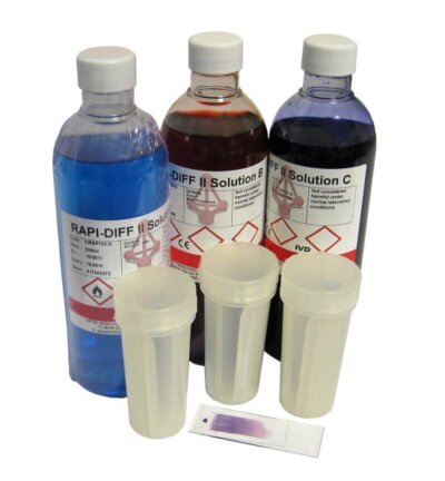
Detecting the possible presence of subclinical Microsporum infection requires a sensitive and reliable test that can be performed on the widest range of appropriate source material including hair roots, dandruff, scabs and skin scrapings. Vetlab Supplies is one specialist who offers Mykodermo Assay Trio, which is an innovative diagnostic test simplifying the detection of the most clinically significant dermatophytosis: Microsporum spp (M.canis, M.gypseum, M.pesicolor), Trichophyton spp (T.verrucosum, T.menta grophytes) and Epidermophyton spp.

Visit Our Website: www.vetlabsupplies.co.uk

Premiere Plans Give Flatter Fields

Premiere Plans Give Flatter Fields

Professional Level Veterinary Microscopes

If you’re a frequent user demanding a high level of performance of your laboratory microscope, ‘Planar’ or ‘Plan’ Objective Lenses give you a clear image that’s in focus at the edges as well as in the centre.

How Your Microscope Works

Microscopes work by deflecting the path of light rays to make objects look many times larger – up to a 1000 times larger – than life. In modern laboratory microscopes, controllable light is provided by the built-in tungsten filament, halogen or LED light source, focussed onto the sample through the substage condenser.

Light passes through the sample or slide mounted on the microscope’s movable stage. Double-layer stages allow the operator to move the sample around in very small increments while vernier calibrated scales let the operator record the position of important findings and locate them again.

Plan Objectives on a Microscope Nosepiece

Bending The Light Fantastic

Light passing through the sample enters the microscope tube at the objective lens. In the pioneer days of microscopes, the objective was a single convex lens resembling a highly polished bead of glass. A single lens will deflect light rays sufficient to effectively magnify the image, but also introduces a number of other unhelpful distortions or ‘aberrations’.

The image created by the objective lens is further magnified by the eyepiece lenses and focussed onto the retina of the operator’s eye. A binocular microscope or ‘stereo-microscope’ allows the operator to view the sample with both eyes.

Relieving Operator Fatigue and Reducing Laboratory Error

One of the most annoying and difficult to correct objective lens aberrations is the loss of ‘flat field’ or ‘planar image distortion’. Uncorrected, an objective lens will create a curved rather than flat image on the retina of the operator’s eye.

This means that while the centre of a sample might be in sharp focus, the edges of the image will be fuzzy and blurred requiring the operator to continually refocus within each field of view in, for instance, a stained blood-smear slide. Constant refocusing can cause operator fatigue, eye strain and inefficient use of laboratory time and even introduce errors.

Planar or Plan Objective Lenses adjust correctly the paths of light rays passing through them to create a flat image that’s in focus at the edges as well as in the centre, reducing eye-strain and operator fatigue.

Choosing Plan Objective Premiere Microscope

Vetlab Supplies’ Premiere 1600 and Premiere 2000 veterinary microscopes feature Infinity Plan Objective Lenses. The benefit of a Plan Objective is perhaps most noticeable when taking photo-micrographs using the trinocular photographic head. These professional quality microscopes also exploit the advances of Revolutionary Infinity Optics, holding the light rays from the objective parallel – as if focussed on infinity – to form a clearer, sharper image in the eye or in a digital camera.

For more information about our great range of microscopes visit our website at www.vetlabsupplies.co.uk or Telephone: 01798 874567

VetCompass Companion Animal Surveillance

VetCompass Companion Animal Surveillance

Where Vets Go When They Need to Know

Evidence-based veterinary testing, diagnosis and treatment depends on the ready availability of reliable information and statistical data. VetCompass, operated and maintained by the University of London’s Royal Veterinary College (RVC) is claimed as the world’s largest merged database of veterinary clinical records.

Risk Factors and Demographics in Companion Animal Disorders

The title VetCompass comes from a contraction of ‘Companion Animal Surveillance System’ a not-for-profit collaboration between the RVC and the University of Sydney. This international initiative aims to collate information and data on the range and frequency of domestic pet health issues. Analysis of the collected data provides veterinary professionals with the tools they need to identify important trends, risk factors and demographics in companion animal disorders.

VetCompass collects clinical and veterinary laboratory data from vets and vet labs in general practice on a day to day basis. The pooled data from a wide range of sources is then merged into an accessible single database making available the vast amount of data that would otherwise remain hidden away in thousands of unconnected local practice records.

Information from Almost 6 Million Animals

VetCompass began as pilot project collating data on the antibiotic and glucocorticoid treatments dispensed to pet animals in a small number of UK veterinary practices. From 2009, the initiative grew to include information from almost 6 million animals. Collaboration with the University of Sydney began in 2013 as ‘VetCompass Australia’. A targeted project, ‘VetCompass Equine’, designed to gather data on ailments and health risks to horses was launched in 2016.

The VetCompass approach to surveillance and data gathering has been applied to diseases ranging through epilepsy, cancer, skin disease, endocrinopathies and heart disease. As well as collating information on the disease, the project also gathers demographic facts and figures on such generalities as longevity and mortality, the occurrence of parasitism on pets and the frequency of accidents in dogs and cats.

Improved Road Safety Awareness

A recent VetCompass survey reported that although only 0.41% of dogs presented at UK vets were victims of road traffic collisions (RTC), almost 25% of those dogs subsequently died or required euthanasia. One demonstration of the benefit of such a large data set is the amount of detail it contains. The survey showed that male dogs were 40% more likely than females to suffer an RTC. Animals aged three years or less were almost 3 times more susceptible to RTC that dogs of 14 years or more. The authors hope that these facts will inform and encourage improved road safety awareness and better streetwise management of dog owners.

Visit our website to see our full range of Veterinary Products, www.vetlabsupplies.co.uk or contact us on 01798 874567.

How to Balance Your Veterinary Centrifuge for Optimal Performance

How to Balance Your Veterinary Centrifuge for Optimal Performance

A reliable, well-balanced benchtop centrifuge is a critical piece of equipment in any busy veterinary diagnostic laboratory. It is pivotal to many veterinary tests, diagnoses and handling procedures while optimising its lifespan and operational safety.

But why is properly balancing your centrifuge before each use so important? How exactly should you go about balancing it?

This guide will walk you through everything you need to know about balancing your veterinary centrifuge for optimal performance, longevity, and safety.

How Does a Balanced Centrifuge Work?

A laboratory centrifuge works by rapidly spinning samples to separate components based on their density. The rotor holds various laboratory samples securely in a fixed position, which, when spun on a vertical spindle, produces strong centrifugal forces – up to several thousand times the force of gravity!

This is known as g-force, which magnifies even tiny differences in weight distribution within the samples and rotor.

Why is It Important to Balance Your Laboratory Centrifuge?

G-forces created by the centrifugation worsen any weight imbalances around the rotor. Even the slightest difference in weight will cause the centrifuge to shake and vibrate uncontrollably, causing undue stress and wear on centrifugal components like motors and bearings. Uneven loads on centrifuge rotors are the most common reason they break down and malfunction.

Over time, this vibrational stress could impact the accuracy of your centrifuge and, in extreme cases, even cause catastrophic failure or explosions. That’s why balancing a high-speed piece of equipment like this is critical.

Follow these simple rules every time you use your centrifuge in your veterinary laboratory or diagnostics:

  • Load the rotor symmetrically – distribute weight evenly on both sides
  • Always use same-size, equally weighted lab samples opposite each other
  • Balance odd samples with matching dummy tubes opposite
  • Keep the inside of the centrifuge chamber clean and dry

In essence, by making sure that each centrifuge is balanced with an even and symmetrical load, it will continue to work optimally with minimal chance of malfunctioning.

We’d recommend checking our visual centrifuge balancing PDF guide below for additional instructions.

How To Balance Your Centrifuge

7 Simple Steps for Balancing Your Lab Centrifuge

Whether you’re centrifuging haematocrit tubes, blood sample tubes, Eppendorf tubes, universal containers or commercial diagnostics such as Ovatube tubes, here are seven simple rules to prolong the life, performance, reliability and safety of your veterinary centrifuge.

  1. Choose the proper rotor for your sample tubes.

Always select a rotor designed specifically for the type and size of consumables you’ll be centrifuging. Using the wrong rotor can increase the chances of accidents or broken tubes.

  1. Inspect the rotor before loading.

Check that the rotor is in good condition with no cracks or damage and make sure it is securely attached to the drive shaft. Wipe the interior clean and dry before loading samples.

  1. Organise samples into matching pairs.

Make sure tubes are the same type and filled to equal levels. Pair each one with a matching tube to place on the opposite side of the rotor.

  1. Load pairs symmetrically into the rotor.

Place each pair directly across from each other, aligned through the central axis. Distribute weight evenly to balance the centrifuge.

  1. Add balance tubes if needed.

If you end up with an odd unpaired tube, make a matching dummy sample with water to act as a balance. Load this opposite the single tube.

  1. Secure the lid and check the balance

Close the lid safely and check that the centrifuge is level. Give it a gentle spin by hand to test if it wobbles before starting the cycle.

  1. Follow safety procedures

Proper balancing of centrifuges only takes a moment and can pay off in a big way. Getting into the habit of checking and balancing regularly will result in safer operation, better performance, and longer equipment lifespan.

Vetlab Supplies provides top centrifuges, tubes, and accessories specially designed for veterinary, clinical, and research applications. Our team is always happy to offer balancing tips and advice to keep your lab running smoothly.

For all your veterinary centrifuge and lab supply needs, contact the experts at Vetlab Supplies on 01798 874567 or visit vetlabsupplies.co.uk.

Online And Social Media Ads Drive ‘Impulse Buy’ Pet Purchases

Online And Social Media Ads Drive ‘Impulse Buy’ Pet Purchases

With Christmas on the way, animal charities and welfare organisations are flagging up the dangers around online and social media pet ads that can lead to impulse buying of very young, unhealthy and misrepresented kittens.

Online classified ads account for a staggering 400,000 dogs and 100,000 cats advertised for sale through online pet shops, breeders and social media. Based on a survey by Blue Cross and the EU Dog and Cat Alliance, 57% of pet purchases were made through online and social media pet ads. Of those surveyed, 95% reported problems with online pet ads ranging from fraudulent ID, banned breeds, false advertising and concerns for the animal’s health.

Lack of the most basic health and welfare information

Complaints included a lack of the most basic health and welfare information. Many of the puppies and kittens advertised were too young to be separated from their mothers and litter-mates. Many were underweight, ill-adjusted to life as a pet, un-chipped and even pregnant.

Most animal welfare experts agree that kittens and pups shouldn’t be separated from their mother and litter-mates before eight weeks old. Many professional breeders won’t allow separation until twelve weeks, permitting a naturally gradual, rather than abrupt, end to nursing. Pups and kittens taken from their litters too soon miss out on the social development vital to a well-adjusted, contented and confident pet.

First Vaccinations for kittens

Kittens should receive their first of their vaccinations against common feline infections, including cat flu, from 8-9 weeks old, preferably before joining a new household. If the new addition is to be a companion to other cats, testing cats for serious diseases such as Feline Leukaemia Virus (FeLV), Feline Immunodeficiency Virus (FIV), Feline Coronavirus (FCoV) and Feline Infectious Peritonitis Virus (FIPV) and might be a really good idea.

First Vaccinations for Puppies 

With pups, vaccinations against distemper, parvovirus, kennel cough, leptospirosis and parainfluenza can begin at 4-6 weeks, with boosters 6-12 months later. Pups don’t have full disease protection until two weeks after completing the course and shouldn’t be exposed to infection risks before then.

Online pet sales in the EU remain largely unregulated. Only Malta obliges dealers to register as online pet shops. UK and Ireland websites are asked to commit the Pet Advertising Advisory Group (PAAG) code, but this is voluntary and, to date, only 6 websites are signed up.

Almost one in six pups experience illness

The Kennel Club reports that for pups purchased in twenty minutes or less, almost one in six experience illness, require ongoing veterinary treatment, or die within their first six months. A rate three times higher than pups chosen over more than an hour of thought and discussion.

The BVA have also expressed concern as to why so many online retailers are apparently ignorant or ignoring the health and welfare of pets. Tackling the issues, says their senior vice president, comes down to 3 things: working with voluntary codes, maintaining pressure to improve the effectiveness of legislation and educating buyers on the responsibilities of pet ownership.

To find out more about our large range of veterinary diagnostic test kits visit our website: www.vetlabsupplies.co.uk or Telephone: 01798 874567

Veterinary Centrifuges: No Longer the Ugly Sister of Laboratory Equipment

Veterinary Centrifuges: No Longer the Ugly Sister of Laboratory Equipment

Maybe it was the challenge of perfecting what goes on inside a bench-top centrifuge that distracted veterinary equipment designers’ from attending to its outward appearance. Maybe, but not anymore.

It might have seemed difficult for designers to get excited about glamorising a piece of laboratory kit that seems to have one simple function – to spin sample tubes very, very fast. But even that’s an over simplification of what a veterinary laboratory demands from a modern centrifuge.

Quiet running with a digital touch-pad control centre

In laboratory centrifuges, power is nothing without control. In specifying the performance criteria of their new VetSpin Duo Veterinary Centrifuge, Vetlab Supplies Ltd not only sourced a smooth and reliable 10,000rpm brushless motor, they integrated its rapid acceleration and quiet running with a digital touch-pad control centre.

Clear, simple and attractive the touch-pad control panel is intuitive even for first-time and infrequent users. The inclusion of programmed, one-touch pre-sets for the most frequent spin applications makes reproducible methodology safe and certain.

With time and bench space at a premium in the busy veterinary practice, the VetSpin Duo satisfies multiple centrifuge requirements in a single machine and without needing to change the centrifuge head between the most frequent laboratory tasks. Thoughtful design has created a single rotor optimised for both micro-haematocrit capillaries and for larger plastic tubes up to 2ml, ending the need to change rotors between applications.

Built-in safety features

Attention to the safety in the veterinary laboratory, wins for VetSpin Duo the coveted CE (Conformité Européene) directive with the assurance that electrical and mechanical components are certified, and built-in safety features comply with the latest national, European and international regulations.

Before the most recent upgrades to centrifuge regulations, the lid-lock fastened when the motor was running under power, but not when power to the motor was turned off. This meant that, although the power was turned off – and the lid unlocked, the rotor was still turning at high speed.

Ensuring the lid cannot be opened while the rotor is spinning, the VetSpin Duo lid lock will not release until the auto-braked rotor has come to a complete standstill. With the auto-brake activated as soon as the power is turned off, deceleration is smooth, quick, quiet and safe.

And what about those unassuming good looks? Well, even the robust; easy to clean outer case is attractive, functional and efficient. Though as always, true beauty is much more than skin-deep.

To find out more about our large range of veterinary products visit our website: www.vetlabsupplies.co.uk or Telephone: 01798 874567

The Light Fantastic – Veterinary Handheld Refractometers

The Light Fantastic – Veterinary Handheld Refractometers

In the veterinary laboratory or out in the field, the handheld veterinary refractometer is a compact and effective diagnostic aid for detecting early indications of diseases including Feline Infectious Peritonitis (FIPV) and Chronic Kidney Disease (CKD).

In other professions, this pocket-sized instrument is used to determine salinity, sugar content, the concentration of industrial coolants, anti-freeze and even the quality of gemstones. In veterinary medicine, the device is readily applied to the measurement of protein in serum, plasma or abdominal fluid, or the specific gravity of a urine sample.

Using a handheld veterinary refractometer is simple

Transparent materials influence the speed and at which light travels through them. Light rays travelling in a low-density medium, such as air, are slowed and so ‘bent’ off course when they enter a denser medium such as water, glass or solution of dissolved solids.  Measuring the angle of bending (refraction) offers a simple means of assessing the density of the medium or the concentration of solids in a solution.

Using a handheld veterinary refractometer is simple.  The sample to be measured is introduced between the flip-up glass cover and the underlying glass prism.  The semi-opaque ground glass cover, which filters and disperses light evenly through the sample acts as a screen against which the internal calibrations scale is displayed.

Ambient light passes through the sample

Ambient light passing through the sample is deflected at a greater or lesser angle according to the density of the sample. Using distilled water as a ‘zero’ indicator, the point at which deflected light illuminates the scale, viewed through the eyepiece, indicates the relative concentration of dissolved solids in the sample.

Refractive index measurement is a quick and practical aid in the diagnosis of animal diseases where abnormally high or low concentrations of protein, salt or other solutes in an animal’s body fluid are key diagnostic symptoms.

Conditions, where body fluid density measurement can have a critical diagnostic role, include the cat diseases Feline Infectious Peritonitis and Chronic Kidney Disease.

Feline Infectious Peritonitis (FIP) is caused by a strain of Feline Coronavirus (FCoV).  Known in the veterinary laboratory Feline Infectious Peritonitis Virus (FIPV), it damages blood vessels (vasculitis) causing fluid to leak into the abdominal and chest cavities.  This fluid has an abnormally high protein concentration which is quickly and easily detected by its high refractive index.  The FASTest diagnostic test kits for FcoV and FIPV will help confirm the diagnosis.

A handheld veterinary refractometer provides a quick and simple tool to help vets diagnose Chronic Kidney Disease. Screening for changes in Urine Specific Gravity (USG), together with serum creatinine measurement, can help older cats live longer by detecting indications of renal disease before clinical symptoms become apparent.

To find out more about our large range of veterinary products visit our website: www.vetlabsupplies.co.uk or Telephone: 01798 874567

Got a Sick Cat? – There’s an App for That!

Got a Sick Cat? – There’s an App for That!

Veterinary apps you can download to your smartphone could mean that home-based veterinary diagnostic testing and even treatment is just a screen-swipe away.

Veterinary smartphone apps could change the way pet owners interact with their veterinary surgery. As apps get smarter, they might even replace visits to the vet altogether.

Vets already use apps to access important records and data out of hours or away from the surgery. They can search online resources ranging from guides for antimicrobial treatments, medical regimens, parasite maps and drug effectiveness and toxicity. There’s also a number of useful dosage calculation apps.

Smartphone technology – already in use

Human medicine already uses smartphone technology. Match the right sensor with the right app, and your smartphone can measure, record and transmit everything from your heart and breathing rate, to your blood oxygen and blood sugar levels.

Smartphone apps really do only one of two things. Either they receive and process information for you, or they process and send out information about you. It doesn’t really matter what that information is. Whether it’s information about you or about your pet, it’s all the same to your phone.

So how long before there’s an app that combines online veterinary resources, your pet’s health history and the sensor-based examination your vet would make at the surgery? And then, why not an app to process all that information and make a diagnosis and prescribe a course of treatment?

All this is technically possible, but only if the interaction between you, your vet and your pet was simply a matter of data processing. It’s a relatively simple programming job to match up all the inputs with an output. But what about the missing inputs?

No app will ever measure how much you care about your pet

What’s missing from ‘veterinary diagnosis by app’ is what you know about your pet, and what your vet knows about you and your pet together. There’s no app that substitutes for this all-important relationship. No app measures how much you care about your pet or your hopes, expectations and even fears.

That’s what Evidence Based Veterinary Medicine (EBVM) is all about. It’s why your vet is ideally placed to examine your pet personally, talk to you about your pet and then help you understand the veterinary laboratory tests, diagnostics and treatments that serve both you the best.

Until there’s an app that lets your cat call up the vet and make the appointment itself, you the pet owner will always be the one with the crucial information your vet needs to give the best diagnosis, treatment and care.

Colour Me Clearly – Biological Stains and Veterinary Microscopy.

Colour Me Clearly – Biological Stains and Veterinary Microscopy.

It’s an unfortunate fact that most things of interest in veterinary microscopy are all more or less colourless. Vetlab’s RAPI-DIFF II microscope slide staining kits help time-strapped veterinary laboratories make the invisible clearly visible.

Practically every living structure at the microscopic level exists in water and consists mostly of water. It’s so much a lack of colour that makes cells and cell structures difficult to see microscopically, it’s a lack of what veterinary microscopists call ‘contrast’.

Poor contrast leaves your subject no brighter or darker

Contrast is what makes an object of interest stand out from its background. In photos and TV images, poor contrast leaves your subject no brighter or darker than the less interesting stuff around, behind or even in front of it.

In a poor contrast picture, you don’t know where to look or even what you’re looking at. Poor contrast means disappointing holiday snaps, but could mean disaster when veterinary diagnosis depends on the micro-exam of a sample from your poorly pet.

Developing biological microscope stains – like dyes used in colour fabrics

Since the invention of microscopes in the 1590s, laboratories have worked to improve the contrast by developing biological microscope ‘stains’. Microscopic stains act just like the dyes used to colour fabrics.

Scientists found that some stains made particular microscopic structures stand out from the less interesting structures around them. Perhaps the two most interesting and important structures in veterinary microscopy are:

  • The Cytoplasm. This is all the stuff that’s inside the cell membrane. Staining the cytoplasm makes the cells stand out and easier to see. It’s also easier to determine the shape of cells which helps diagnose some parasitic infections
  • The Nucleus. This is the cell’s central control unit that contains the DNA. Many cells, especially infection-fighting white blood cells, have particular shaped nuclei. Identifying white cell types can help vets diagnose some diseases.

In 1891 the Russian physician Dmitri Leonidovich Romanowsky invented a balance of microscope stains for cell cytoplasm and nuclei that not only made cells easier to find and examine, but actually helped identify different kinds of cell.

Romanowsky’s proven staining principle

Vetlab’s RAPI-DIFF II kit incorporates Romanowsky’s proven staining principle into a complete, convenient and simple to use microscope slide staining kit.

Pre-prepared solutions create a simple three–dip process that fixes the sample to the microscope slide, stains cell cytoplasm with a Thiazine-Eosin Y, and contrasts clearly with the Methylene Blue stained nuclei. Selectively rinsing with pH buffered solution or further dipping gives veterinary labs a practical tool for recognising and differentiating a range of cell types and diagnosing abnormalities.

Fire Up The Vet Lab Response Vehicle! – Equipping a Mobile Veterinary Laboratory

Fire Up The Vet Lab Response Vehicle! – Equipping a Mobile Veterinary Laboratory

Planning a mobile veterinary laboratory is less about giving the right answers and more about making sure you’ve asked the right questions.

Whatever you can do in a mobile test lab, you can do better in a well equipped, purpose-built veterinary laboratory. So the first question is, why would you want a mobile lab anyway?

The only reason for taking a mobile lab to animals in the field – often literally, is because the animals can’t come to you. That could be because the animals are too ill, too far away, too many or because the outcome of a laboratory diagnostic enables an immediate veterinary response without a second on-site visit.

Justifying the investment in a mobile resource

The next question might be what could you usefully do on-site? This will depend on your vet lab’s customer base. With a large animal practice, screening large numbers of cattle, sheep, poultry and other livestock for parasites and blood disorders might justify the investment in a mobile resource. Practices with a mainly small animal base might usefully offer on-site veterinary diagnostics to kennels, catteries and breeders where numbers create an economy of scale.

With a clear idea of why you need a mobile vet lab and what you want to do, you can start to think about what you need to put into it. The first requirement is simply space. If you can’t transport enough space to work in – either self-propelled or towed, then you might consider simply transporting equipment and setting up a lab in-situ.

Going truly mobile 

In any situation, don’t overlook the basics. You’ll need solid, stable work surfaces that you can keep clean. You’ll need enough lighting to see what you’re doing, and you’ll need to keep warm… or cool. Electricity is essential, as is a supply of water and safe drainage or containment of waste. If you’re going truly mobile, you may be surprised by the amount of space you need for laboratory consumables – even before you consider how to squeeze in cold or frozen storage.

Before committing to your vehicle, trailer or even tent, lay out a floor plan and mark out where you’d put your lab benches, storage and veterinary equipment including centrifuges, microscopes and small equipment. Then ask… do I really need to do this at all?

Many of the most essential veterinary diagnostic kits for the most frequently required tests need no laboratory facilities or equipment. Vetlab Supplies Ltd`s FASTest range of diagnostic kits are quick, simple to use, reliable and with long shelf-lives even at room temperature. With FASTest veterinary diagnostics, you can turn your car, your field-kit, a bicycle or even your jacket pockets into a fully-functional, quick response veterinary test laboratory.